. 2011 Mar 17;471(7338):368-72.
doi: 10.1038/nature09857. Epub 2011 Mar 2.

RIP3 mediates the embryonic lethality of caspase-8-deficient mice

Affiliations

RIP3 mediates the embryonic lethality of caspase-8-deficient mice

William J Kaiser et al. Nature. .

Abstract

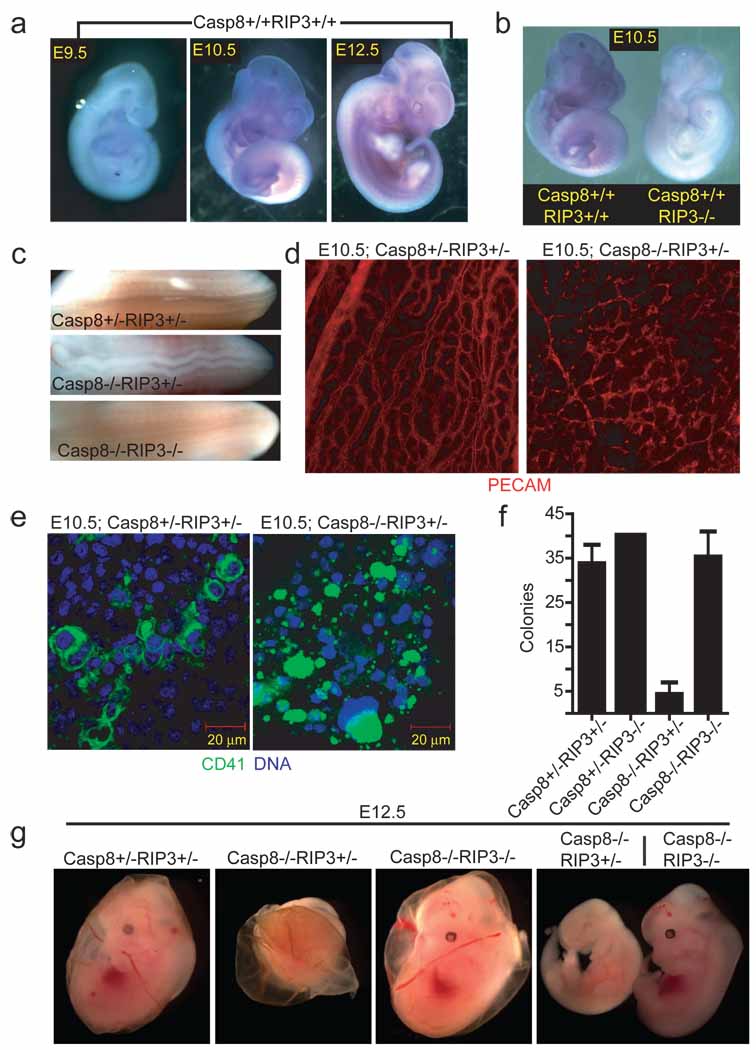
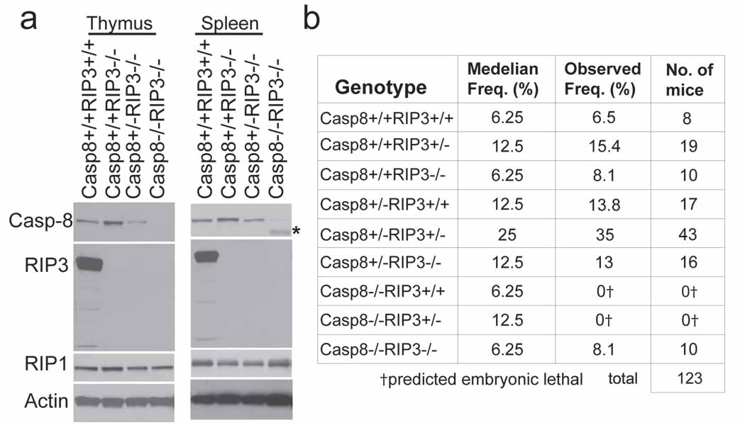
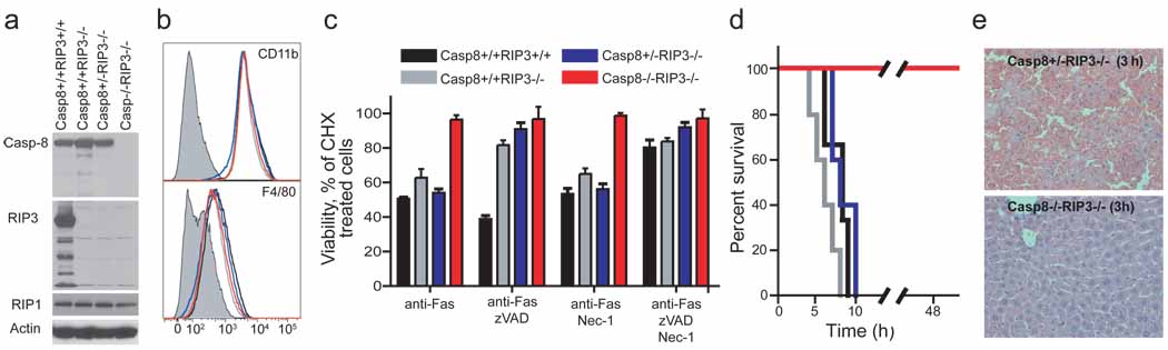
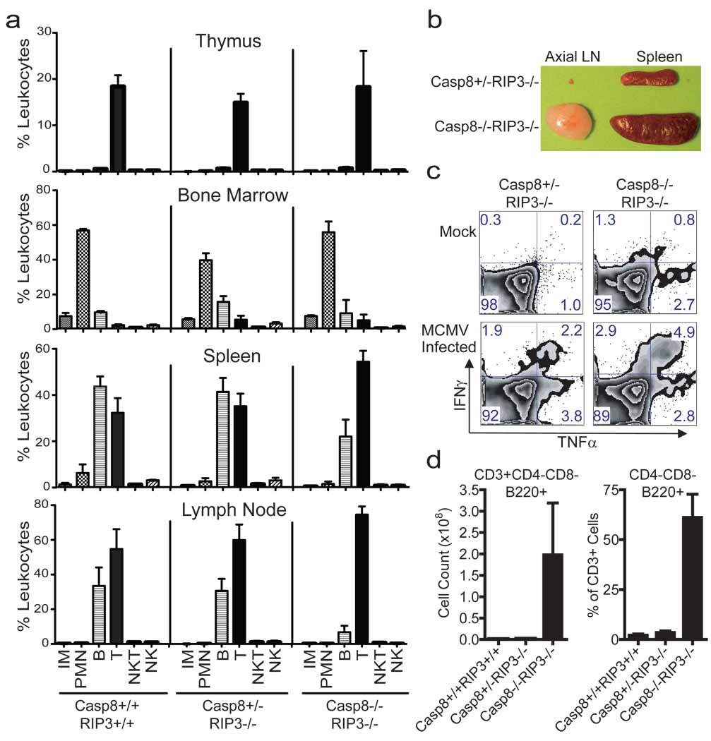
Apoptosis and necroptosis are complementary pathways controlled by common signalling adaptors, kinases and proteases; among these, caspase-8 (Casp8) is critical for death receptor-induced apoptosis. This caspase has also been implicated in non-apoptotic pathways that regulate Fas-associated via death domain (FADD)-dependent signalling and other less defined biological processes as diverse as innate immune signalling and myeloid or lymphoid differentiation patterns. Casp8 suppresses RIP3-RIP1 (also known as RIPK3-RIPK1) kinase complex-dependent necroptosis that follows death receptor activation as well as a RIP3-dependent, RIP1-independent necrotic pathway that has emerged as a host defence mechanism against murine cytomegalovirus. Disruption of Casp8 expression leads to embryonic lethality in mice between embryonic days 10.5 and 11.5 (ref. 7). Thus, Casp8 may naturally hold alternative RIP3-dependent death pathways in check in addition to promoting apoptosis. We find that RIP3 is responsible for the mid-gestational death of Casp8-deficient embryos. Remarkably, Casp8(-/-)Rip3(-/-) double mutant mice are viable and mature into fertile adults with a full immune complement of myeloid and lymphoid cell types. These mice seem immunocompetent but develop lymphadenopathy by four months of age marked by accumulation of abnormal T cells in the periphery, a phenotype reminiscent of mice with Fas-deficiency (lpr/lpr; also known as Fas). Thus, Casp8 contributes to homeostatic control in the adult immune system; however, RIP3 and Casp8 are together completely dispensable for mammalian development.


Figures

Figure 1
Figure 1
Embryonic expression of Rip3. (a) Whole-mount Rip3 in situ hybridization of Casp8+/+Rip3+/+ E9.5 (left panel), E10.5 (middle panel), and E12.5 (right panel) embryos. (b) Whole-mount Rip3 in situ hybridization of Casp8+/+Rip3+/+ and Casp8+/+Rip3−/− E10.5 embryos demonstrating specificity of the probe. (c) View of the neural tube of E11.5 embryos with the indicated genotype. (d) PECAM-1 (CD31) staining of a whole-mount E10.5 yolk sac from a representative Casp8+/−Rip3+/− (left panel) and Casp8−/−Rip3+/− (right panel) embryo (100X). (e) CD41 (green) and nuclear DNA (blue) staining of a yolk sac from a E10.5 Casp8+/−Rip3+/− (left panel) and a Casp8−/−Rip3+/− (right panel) embryo. (f) Numbers of colony-forming cells (CFC) following culture of disrupted E10.5 yolk sacs of the indicated genotype. (g) Photographs of E12.5 embryos and yolk sacs with the indicated genotype. The right panel shows side by side embryos with yolk sacs removed.
Figure 2
Figure 2
Casp8−/−Rip3−/− mice are viable. (a) IB of Casp8, RIP3, RIP1 and β-actin from thymus (left panel) and spleen (right panel). The asterisk denotes elevated heavy IgG heavy chain reactive with secondary antibody in DKO sample. (b) Epistatic analysis of mice born following Casp8+/−Rip3+/− intercross with predicted and observed frequencies.
Figure 3
Figure 3
Sensitivity to DR-induced apoptosis, necroptosis and disease. (a) IB of Casp8, RIP3, RIP1 and β-actin in BMDM derived from mice with the indicated genotype. (b) Relative cell surface expression levels of CD11b (top panel) or F4/80 (bottom panel) shown by red line (DKO), black line (Casp8+/+Rip3+/+), grey line (Casp8+/+Rip3−/−), or blue line (Casp8+/−Rip3−/−) on BMDM stained cells. Isotype control is shown by shaded histogram. (c) Viability of BMDM cultured in CHX (5 µg/mL) and treated with anti-Fas antibody for 18 h in the presence or absence of the caspase inhibitor zVAD-fmk (25 µM) and/or Nec-1 (30 µM). Cell viability was determined by measuring intracellular ATP levels with a Cell Titer-Glo Luminescent Cell Viability Assay kit. Error bars, SD. (n = 4). (d) Kaplan-Meier survival plot of 12 week old Casp8+/+Rip3+/+, Casp8+/+Rip3−/−, Casp8+/−Rip3−/−, and DKO mice injected intraperitoneally with 12.5 µg of anti-Fas Jo-2 antibody. Legend genotypes and line color is the same as in (c). (e) Histology of liver sections from Casp8+/−Rip3−/− (left panel) and Casp8−/−Rip3−/− mice (right panel) 3 h following injection with Jo-2 antibody.
Figure 4
Figure 4
Immune compartment of 16-week-old DKO mice. (a) Live cells from thymus (top panels), bone marrow (second set of panels), spleen (third set of panels) and LN (bottom panels), gated based on forward and side scatter properties, and stained for surface expression of CD19, CD3, CD49b, Ly6C and CD11b to define non-overlapping leukocyte (CD45+) populations. The average and SD for three wild-type (left panels), four Casp8+/−RIP3−/− (middle panels) littermate control and three DKO (right panels) mice showing levels of inflammatory monocytes (IM), polymorphonuclear leukocytes (PMN), B cells, T cells, NK cells and NKT cells. (b) Photograph of the axial LN and spleen from representative mice of the indicated genotype. (c) Flow cytometric analysis of IFNγ and TNFα on T cells from naïve or MCMV-infected (7 days) mice following stimulation with CD3/CD28 antibodies. CD8+ T cells from one representative animal per experimental group are shown. (d) B220 expression was assessed on CD4CD8 splenic T cells. Error bars, SD.

Comment in

References

    1. Strasser A, Jost PJ, Nagata S. The many roles of FAS receptor signaling in the immune system. Immunity. 2009;30:180–192. - PMC - PubMed
    1. Cho YS, et al. Phosphorylation-driven assembly of the RIP1-RIP3 complex regulates programmed necrosis and virus-induced inflammation. Cell. 2009;137:1112–1123. - PMC - PubMed
    1. He S, et al. Receptor interacting protein kinase-3 determines cellular necrotic response to TNF-alpha. Cell. 2009;137:1100–1111. - PubMed
    1. Zhang DW, et al. RIP3, an Energy Metabolism Regulator that Switches TNF-Induced Cell Death from Apoptosis to Necrosis. Science. 2009 - PubMed
    1. Holler N, et al. Fas triggers an alternative, caspase-8-independent cell death pathway using the kinase RIP as effector molecule. Nat Immunol. 2000;1:489–495. - PubMed

Publication types

MeSH terms