. 2010 Nov 4;1(11):e92.
doi: 10.1038/cddis.2010.60.

Glucocorticoids induce long-lasting effects in neural stem cells resulting in senescence-related alterations

Affiliations

Glucocorticoids induce long-lasting effects in neural stem cells resulting in senescence-related alterations

R Bose et al. Cell Death Dis. .

Abstract

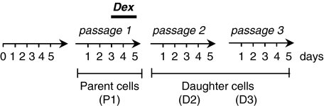
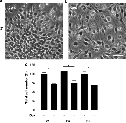
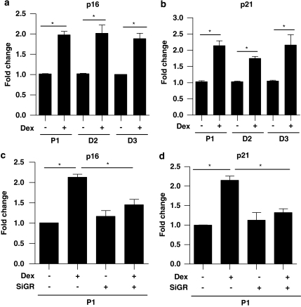
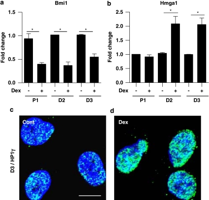
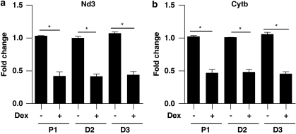
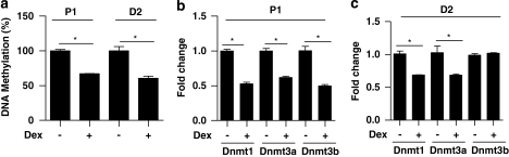
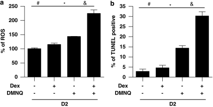
Alterations in intrauterine programming occurring during critical periods of development have adverse consequences for whole-organ systems or individual tissue functions in later life. In this paper, we show that rat embryonic neural stem cells (NSCs) exposed to the synthetic glucocorticoid dexamethasone (Dex) undergo heritable alterations, possibly through epigenetic mechanisms. Exposure to Dex results in decreased NSC proliferation, with no effects on survival or differentiation, and changes in the expression of genes associated with cellular senescence and mitochondrial functions. Dex upregulates cell cycle-related genes p16 and p21 in a glucocorticoid receptor(GR)-dependent manner. The senescence-associated markers high mobility group (Hmg) A1 and heterochromatin protein 1 (HP1) are also upregulated in Dex-exposed NSCs, whereas Bmi1 (polycomb ring finger oncogene) and mitochondrial genes Nd3 (NADH dehydrogenase 3) and Cytb (cytochrome b) are downregulated. The concomitant decrease in global DNA methylation and DNA methyltransferases (Dnmts) suggests the occurrence of epigenetic changes. All these features are retained in daughter NSCs (never directly exposed to Dex) and are associated with a higher susceptibility to oxidative stress, as shown by the increased occurrence of apoptotic cell death on exposure to the redox-cycling reactive oxygen species (ROS) generator 2,3-dimethoxy-1-naphthoquinone (DMNQ). Our study provides novel evidence for programming effects induced by glucocorticoids (GCs) on NSCs and supports the idea that fetal exposure to endogenous or exogenous GCs is likely to result in long-term consequences that may predispose to neurodevelopmental and/or neurodegenerative disorders.


Figures

Figure 1
Figure 1
Experimental model. After cortice dissection and cell plating, NSCs were cultured for 5 days before passaging in order to obtain parent cells (P1). After 3 days in culture, P1 cells were exposed to Dex (1 μM) for 48 h. To investigate the long-lasting (heritable) effects of Dex, P1 NSCs were passaged to get daughter cells (D). D2=daughter NSCs from passage 2; D3=daughter NSCs from passage 3
Figure 2
Figure 2
Dex exposure decreases NSC proliferation. Phase contrast images of control (a) and Dex-exposed (1 μM Dex for 48 h) (b) P1 NSCs. Scale bar=50 μm. (c) Total cell number (expressed as percentage from cell counts of control cells) of parent (P1) and D2 and D3 cells. Values are shown as mean±S.E.M. *P<0.05
Figure 3
Figure 3
Dex decreases the number of Ki67-positive NSCs without changing the percentage of nestin-positive cells. Micrographs show Ki67-positive cells (green colour) (a, b) and the percentage of positive cells is given in (c). Cytoplasmatic staining for nestin (green colour) in control (d) and Dex (e) NSCs. Nuclei are stained with Hoechst 33342 (blue colour). Values are shown as mean±S.E.M. *P<0.05. Scale bar=50 μm
Figure 4
Figure 4
The long-lasting upregulation of p16 and p21 expression is GR dependent. Gene expression analyses by q-PCR showed that p16 (a) and p21 (b) genes were upregulated in P1 NSCs (immediately after 48 h Dex exposure), as well as in D2 and D3 NSCs (24 h after the respective passaging). siRNA nucleofection to knock down GR blocked the Dex-induced p16 (c) and p21 (d) upregulation. Relative quantification expression values were calculated by the arithmetic equation 2−ΔΔCT. The amount of target genes was normalized to Hprt and the relative increase was equal to 2−Δ(ΔCTDex–ΔCTcontrol). *P<0.05
Figure 5
Figure 5
Dex induces the expression of senescence markers in NSCs. q-PCR analyses showed that the expression level of Bmi1 (a) was significantly decreased in P1 NSCs 48 h after Dex exposure, as well as in D2 and D3 NSCs 24 h after the respective passaging. The increased expression of Hmga1 mRNA (b) was observed only in D2 and D3 cells. Confocal images of control (c) and Dex (d) NSCs showing an enrichment of heterochromatin protein 1 gamma (HP1γ) (green colour) induced by Dex. Nuclei were counterstained with Hoechst 33342 (blue colour). *P<0.05, Scale bar=50 μm
Figure 6
Figure 6
Dex alters the expression of mitochondrial genes. A long-lasting significant decrease of Nd3 (a) and Cytb (b) mRNA expression was induced by Dex in NSCs. Values are shown as mean±S.E.M. *P<0.05
Figure 7
Figure 7
Dex modifies global DNA methylation. Global levels of cytosine methylation were decreased in P1 and D2 Dex-exposed NSCs (a) with concomitant downregulation of methyltransferases Dnmt1 and Dnmt3a mRNA (b and c). The expression level of Dnmt3b mRNA was downregulated only in P1 cells (b). Values are shown as mean±S.E.M. *P<0.05
Figure 8
Figure 8
Dex increases NSC susceptibility to oxidative stress. D2 cells exposed to the oxidative stress inducer DMNQ for 8 h showed a significant increase in ROS accumulation (a) and an increased number of TUNEL-positive apoptotic cells, 24 h after DMNQ exposure (b). Values are shown as mean±S.E.M. *P<0.05

References

    1. Fowden AL, Forhead AJ. Endocrine mechanisms of intrauterine programming. Reproduction. 2004;127:515–526. - PubMed
    1. Barker DJ. Outcome of low birth weight. Horm Res. 1994;42:223–230. - PubMed
    1. Strang-Karlsson S, Räikkonen K, Pesonen AK, Kajantie E, Paavonen EJ, Lahti J, et al. Very low birth weight and behavioral symptoms of attention deficit hyperactivity disorder in young adulthood: the Helsinki study of very-low-birth-weight adults. Am J Psychiatry. 2008;165:1345–1353. - PubMed
    1. Räikkönen K, Pesonen AK, Heinonen K, Kajantie E, Hovi P, Jarvenpaa AL, et al. Depression in young adults with very low birth weight: the Helsinki study of very low-birth-weight adults. Arch Gen Psychiatry. 2008;65:290–296. - PubMed
    1. Seckl JR. Prenatal glucocorticoids and long-term programming. Eur J Endocrinol. 2004;151 (Suppl 3:U49–U62. - PubMed

Publication types

MeSH terms