. 2011 Feb 14;19(4):3657-66.
doi: 10.1364/OE.19.003657.

Real-time bulk-motion-correction free Doppler variance optical coherence tomography for choroidal capillary vasculature imaging

Affiliations

Real-time bulk-motion-correction free Doppler variance optical coherence tomography for choroidal capillary vasculature imaging

Gangjun Liu et al. Opt Express. .

Abstract

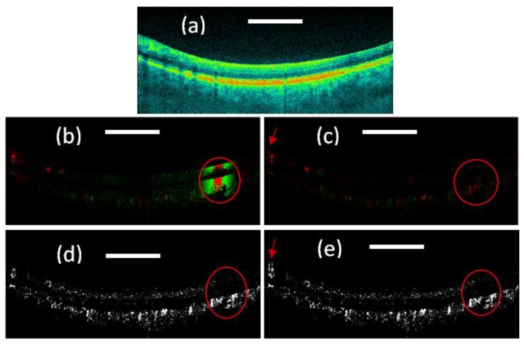
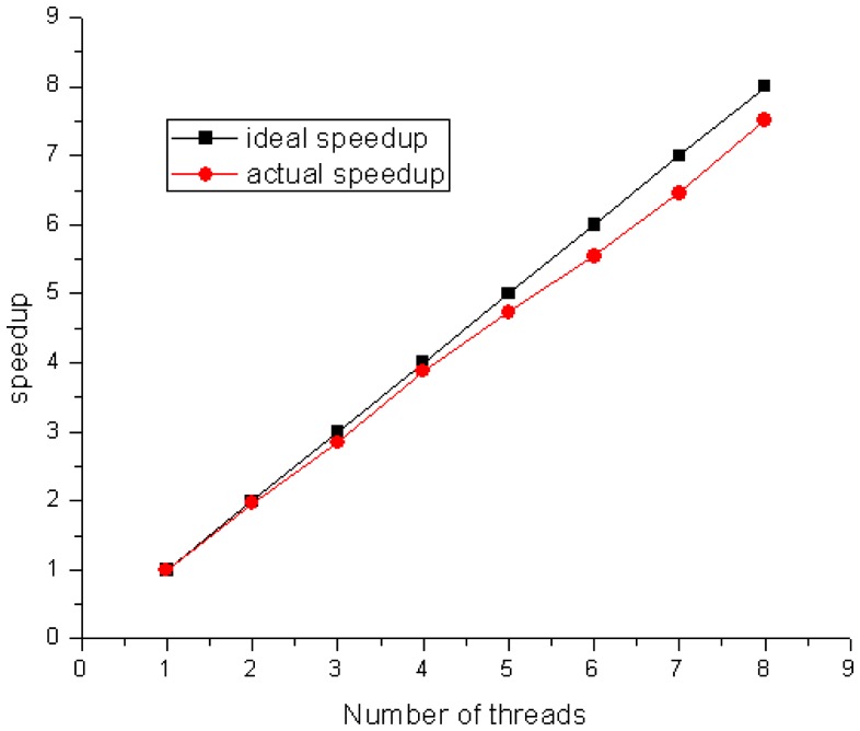
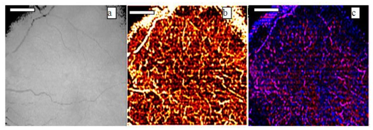
In this paper, we analyze the retinal and choroidal blood vasculature in the posterior segment of the human eye with optimized color Doppler and Doppler variance optical coherence tomography. Depth-resolved structure, color Doppler and Doppler variance images are compared. Blood vessels down to the capillary level were detected and visualized with the optimized optical coherence color Doppler and Doppler variance method. For in-vivo imaging of human eyes, bulk-motion induced bulk phase must be identified and removed before using the color Doppler method. It was found that the Doppler variance method is not sensitive to bulk-motion and the method can be used without correcting the bulk-motion when the sample-movement-induced velocity changes gradually. Real-time processing and displaying of the structure and blood vessel images are very interesting and is demonstrated using a dual quad-core Central Processing Unit (CPU) workstation. High resolution images of choroidal capillary of the vasculature network with phased-resolved color Doppler and Doppler variance are shown.


Figures

Fig. 1
Fig. 1
(a) OCT structure image; (b) Color Doppler OCT image without bulk motion correction; (c). Color Doppler OCT image with bulk motion correction; (e) Doppler variance OCT image without bulk motion correction; (f) Doppler variance OCT image with bulk motion correction. Scale bar: 1mm.
Fig. 2
Fig. 2
Speed-up curve for a workstation with dual quad-core CPUs of Intel Xeon processors X5550.
Fig. 3
Fig. 3
(a)OCT projection, (b) Doppler variance projection and (c) color Doppler projection images of choroidal vessels. Scale bar: 1mm.

References

    1. Huang D., Swanson E. A., Lin C. P., Schuman J. S., Stinson W. G., Chang W., Hee M. R., Flotte T., Gregory K., Puliafito C. A., Fujimoto J. G., “Optical coherence tomography,” Science 254(5035), 1178–1181 (1991).10.1126/science.1957169 - DOI - PMC - PubMed
    1. Chen Z., Milner T. E., Dave D., Nelson J. S., “Optical Doppler tomographic imaging of fluid flow velocity in highly scattering media,” Opt. Lett. 22(1), 64–66 (1997).10.1364/OL.22.000064 - DOI - PubMed
    1. Chen Z., Milner T. E., Srinivas S., Wang X. J., Malekafzali A., van Gemert M. J. C., Nelson J. S., “Noninvasive imaging of in vivo blood flow velocity using optical Doppler tomography,” Opt. Lett. 22(14), 1119–1121 (1997).10.1364/OL.22.001119 - DOI - PubMed
    1. Izatt J. A., Kulkarni M. D., Yazdanfar S., Barton J. K., Welch A. J., “ In vivo bidirectional color Doppler flow imaging of picoliter blood volumes using optical coherence tomography,” Opt. Lett. 22(18), 1439–1441 (1997).10.1364/OL.22.001439 - DOI - PubMed
    1. Zhao Y., Chen Z., Saxer C., Shen Q., Xiang S., de Boer J. F., Nelson J. S., “Doppler standard deviation imaging for clinical monitoring of in vivo human skin blood flow,” Opt. Lett. 25(18), 1358–1360 (2000).10.1364/OL.25.001358 - DOI - PubMed

Publication types