Review
. 2011 Jun;21(2):102-18.
doi: 10.1007/s11065-011-9163-0. Epub 2011 Mar 3.

Imaging the impact of prenatal alcohol exposure on the structure of the developing human brain

Affiliations
Review

Imaging the impact of prenatal alcohol exposure on the structure of the developing human brain

Catherine Lebel et al. Neuropsychol Rev. 2011 Jun.

Abstract

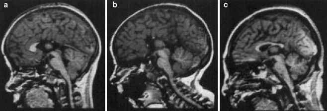
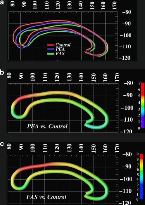
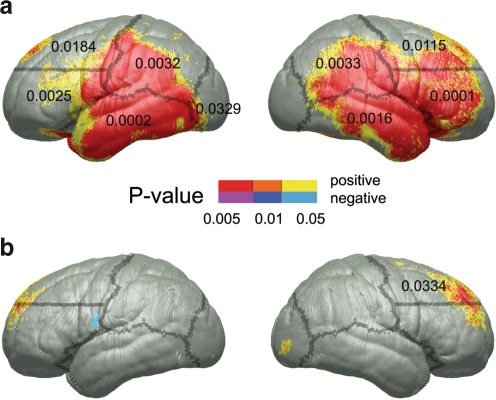
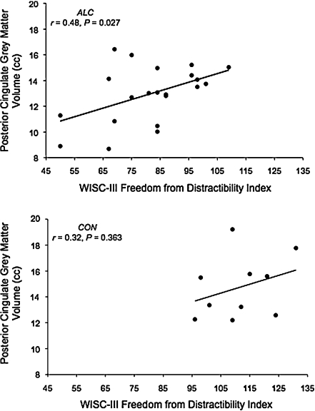
Prenatal alcohol exposure has numerous effects on the developing brain, including damage to selective brain structure. We review structural magnetic resonance imaging (MRI) studies of brain abnormalities in subjects prenatally exposed to alcohol. The most common findings include reduced brain volume and malformations of the corpus callosum. Advanced methods have been able to detect shape, thickness and displacement changes throughout multiple brain regions. The teratogenic effects of alcohol appear to be widespread, affecting almost the entire brain. The only region that appears to be relatively spared is the occipital lobe. More recent studies have linked cognition to the underlying brain structure in alcohol-exposed subjects, and several report patterns in the severity of brain damage as it relates to facial dysmorphology or to extent of alcohol exposure. Future studies exploring relationships between brain structure, cognitive measures, dysmorphology, age, and other variables will be valuable for further comprehending the vast effects of prenatal alcohol exposure and for evaluating possible interventions.


Figures

Fig. 1
Fig. 1
Examples of corpus callosum abnormalities in alcohol-exposed subjects, including (a, b) partial agenesis and (c) hypoplasia. Reproduced from Johnson et al., 1997 with permission. These severe malformations are observed in rare cases of subjects with very heavy prenatal alcohol exposure; however, more subtle callosal abnormalities have been observed in most studies of prenatal alcohol exposure
Fig. 2
Fig. 2
Sowell et al. (2001a), demonstrated changes in corpus callosum displacement. Here, the top row (a) shows callosal shape averages in native space in controls (red), subjects with fetal alcohol syndrome (FAS; green), and subjects with prenatal exposure to alcohol (PEA; blue). The middle row (b) shows displacement differences in millimeters between subjects with PEA and controls; the bottom row (c) shows the differences between FAS subjects and controls. Note that the abnormalities relative to controls that were observed in subjects with FAS and PEA are similar, although they are slightly more extensive in the FAS cohort. The callosal displacement here was significantly related to performance on the California Verbal Learning Test for children (CVLT-C). Figure used with permission
Fig. 3
Fig. 3
Sowell et al. (2008) demonstrated thicker cortices in alcohol-exposed subjects compared to healthy controls. The top row (a) shows the location of cortical thickness differences between exposed subjects and controls and the significance of those changes. They also observed correlations between the cortical thickness and performance on the CVLT-C, a test of verbal learning. The significant correlations were located in the right frontal lobe in exposed subjects (b). Figure used with permission
Fig. 4
Fig. 4
Bjorkquist et al. (2010) found a significant correlation between posterior cingulate gray matter volume and the freedom from distractibility index, indicating that smaller volumes were associated with more distractibility. Figure used with permission
Fig. 5
Fig. 5
Nardelli et al. (2011) observed significantly smaller volume of six deep gray matter structures in alcohol-exposed subjects compared to controls. They also observed significant age-related changes within the globus pallidus (dark blue) for both groups, and within the thalamus (green) for controls (both shown above). The other four structures—the caudate (light blue), putamen, hippocampus, and amygdala (not shown) had no significant age-related changes. Across the age range, the mean volume of exposed subjects remained significantly below that of controls, suggesting consistency across the age range
Fig. 6
Fig. 6
Roussotte et al. (2011) showed correlations between lipometer score (a measure of philtrum dysmorphology) and volume of deep gray matter structures within subjects with prenatal alcohol exposure, demonstrating significantly smaller left pallidum and bilateral thalamus volumes (and a trend in right putamen) in FASD subjects with more severe facial abnormalities. Figure used with permission

References

    1. Abel EL. Prenatal effects of alcohol. Drug and Alcohol Dependence. 1984;14(1):1–10. doi: 10.1016/0376-8716(84)90012-7. - DOI - PubMed
    1. Abel EL. Prenatal effects of alcohol on growth: a brief overview. Federation Proceedings. 1985;44(7):2318–2322. - PubMed
    1. Arango C, Moreno C, Martinez S, Parellada M, Desco M, Moreno D, et al. Longitudinal brain changes in early-onset psychosis. Schizophrenia Bulletin. 2008;34(2):341–353. doi: 10.1093/schbul/sbm157. - DOI - PMC - PubMed
    1. Archibald SL, Fennema-Notestine C, Gamst A, Riley EP, Mattson SN, Jernigan TL. Brain dysmorphology in individuals with severe prenatal alcohol exposure. Developmental Medicine and Child Neurology. 2001;43(3):148–154. - PubMed
    1. Astley SJ. Diagnositc guide for fetal alcohol spectrum disorders: the 4-digit diagnostic code. Seattle: Fetal Alcohol Syndrome Diagnostic and Prevention Network, University of Washington; 2004.