

doi: 10.1002/anie.201007626.
Epub 2011 Mar 2.
The binding of fluorophores to proteins depends on the cellular environment
Affiliations
- PMID: 21370369
- PMCID: PMC4758120
- DOI: 10.1002/anie.201007626
Item in Clipboard
The binding of fluorophores to proteins depends on the cellular environment
Angew Chem Int Ed Engl.
.
No abstract available
Figures
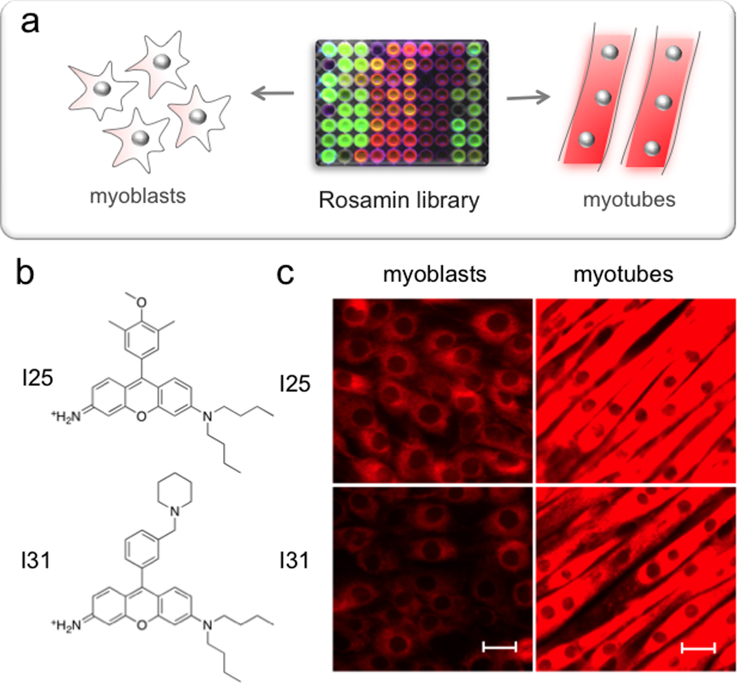
(a) Screen of rosamine library. Myoblasts or myotubes were incubated with 500 nM of library compounds for 2 hours before imaged (b) Chemical structures of selected probes, I25 and I31. (c) Fluorescent images of I25 and I31 before (right) and after (left) muscle differentiation. Scale bar = 20 µm.
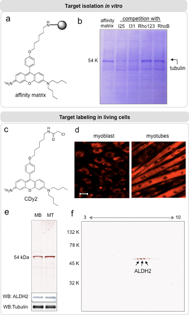
(a) Chemical structure of the affinity matrix used to isolate cellular proteins. (b) SDS-PAGE analysis of bead-bound proteins from C2C12 myotubes. In a competition assay, myotube lysates were pre-incubated with 100 µM of I25, I31, or control compounds (rhodamine 123 and rhodamine B) before the affinity pull-down experiment. (c) Chemical structure of CDy2 for labeling target protein in living cells. (d) Myoblasts (MB) or myotubes (MT) were incubated with CDy2 (500 nM) for 30 min and imaged with a fluorescent microscope. Then, cells were lysed for in-gel fluorescence analysis (λex = 530 nm, λex = 580 nm) (e). (f) 2D-gel analysis for the identification of labeled-protein.
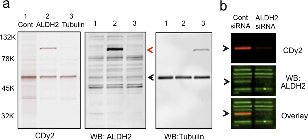
(a) GFP-tagged ALDH2 or tubulin constructs were transfected into HEK293 cells. After 48 hours, cells were labeled with CDy2 and subjected to in-gel fluorescence imaging. Immunoblot analysis shows the endogenous (black arrow) and over-expressed (red arrow) proteins. (b) C2C12 myoblasts were transfected with siRNA against ALDH2. After 72 hours of transfection, cells were labeled with CDy2. Fluorescence labeling patterns of CDy2 were directly compared to ALDH2 immunofluororescence (green).
Similar articles
-
Site-selective labeling at Cys302 of aldehyde dehydrogenase unveils a selective mitochondrial stain.Mol Biosyst. 2011 Aug;7(8):2375-8. doi: 10.1039/c1mb05137g. Epub 2011 Jun 9. Mol Biosyst. 2011. PMID: 21660325
-
An original class of small sized molecules as versatile fluorescent probes for cellular imaging.Chem Commun (Camb). 2019 Jul 2;55(54):7776-7779. doi: 10.1039/c9cc03765a. Chem Commun (Camb). 2019. PMID: 31210218
-
Capture and visualization of hydrogen sulfide by a fluorescent probe.Angew Chem Int Ed Engl. 2011 Oct 24;50(44):10327-9. doi: 10.1002/anie.201104305. Epub 2011 Sep 6. Angew Chem Int Ed Engl. 2011. PMID: 21898737 Free PMC article. No abstract available.
-
[Aldehyde dehydrogenase (ALDH)].Nihon Rinsho. 1997 Feb;55 Suppl:35-9. Nihon Rinsho. 1997. PMID: 9078705 Review. Japanese. No abstract available.
-
Fluorescence spectroscopic methods to analyze drug-tubulin interactions.Methods Cell Biol. 2010;95:301-29. doi: 10.1016/S0091-679X(10)95017-6. Methods Cell Biol. 2010. PMID: 20466142 Review.
Cited by
-
Hot mitochondria?PLoS Biol. 2018 Jan 25;16(1):e2005113. doi: 10.1371/journal.pbio.2005113. eCollection 2018 Jan. PLoS Biol. 2018. PMID: 29370159 Free PMC article.
-
A sensitive mitochondrial thermometry 2.0 and the availability of thermogenic capacity of brown adipocyte.Front Physiol. 2022 Aug 24;13:977431. doi: 10.3389/fphys.2022.977431. eCollection 2022. Front Physiol. 2022. PMID: 36091398 Free PMC article.
-
Regulation of mitochondrial temperature in health and disease.Pflugers Arch. 2022 Oct;474(10):1043-1051. doi: 10.1007/s00424-022-02719-2. Epub 2022 Jul 2. Pflugers Arch. 2022. PMID: 35780250 Free PMC article. Review.
-
Neural stem cell specific fluorescent chemical probe binding to FABP7.Proc Natl Acad Sci U S A. 2012 Jun 26;109(26):10214-7. doi: 10.1073/pnas.1200817109. Epub 2012 Jun 11. Proc Natl Acad Sci U S A. 2012. PMID: 22689954 Free PMC article.
-
Synthesis of Rhodamines and Rosamines Using 3,6-Difluoroxanthone as a Common Intermediate.J Org Chem. 2021 Dec 17;86(24):17856-17865. doi: 10.1021/acs.joc.1c02135. Epub 2021 Nov 24. J Org Chem. 2021. PMID: 34816717 Free PMC article.
References
-
- Giepmans BN, Adams SR, Ellisman MH, Tsien RY. Science. 2006;312:217–224. - PubMed
-
- Wang S, Chang YT. J. Am. Chem. Soc. 2006;128:10380–10381. - PubMed
-
- Lee JW, Jung M, Rosania GR, Chang YT. Chem. Commun. 2003:1852–1853. - PubMed
-
- Li Q, Kim Y, Namm J, Kulkarni A, Rosania GR, Ahn YH, Chang YT. Chem. Biol. 2006;13:615–623. - PubMed
Publication types
MeSH terms
Substances
Grants and funding
LinkOut - more resources
Full Text Sources
Other Literature Sources
