Review
. 2011 Feb;121(2):468-74.
doi: 10.1172/jci44595.

Epithelial-mesenchymal transition (EMT) in kidney fibrosis: fact or fantasy?

Affiliations
Review

Epithelial-mesenchymal transition (EMT) in kidney fibrosis: fact or fantasy?

Wilhelm Kriz et al. J Clin Invest. 2011 Feb.

Abstract

Epithelial-mesenchymal transition (EMT) has become widely accepted as a mechanism by which injured renal tubular cells transform into mesenchymal cells that contribute to the development of fibrosis in chronic renal failure. However, an increasing number of studies raise doubts about the existence of this process in vivo. Herein, we review and summarize both sides of this debate, but it is our view that unequivocal evidence supporting EMT as an in vivo process in kidney fibrosis is lacking.


Figures

Figure 1
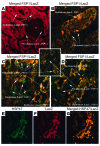
Figure 1. In the kidney, FSP1/S100A4 does not stain fibroblasts or myofibroblast.
(A) Double immunofluorescence for S100A4 (red channel) and ecto-5′nucleotidase (5′NT, green channel) and (B) for S100A4 (red channel) and α-SMA (green channel); nuclei in B are stained by DAPI. (A) 3-mm-thick cryostat section of renal cortex (rat); the thick arrow points to a 5′NT-positive interstitial fibroblast; S100A4-positive cells (thin arrows) are seen inside capillaries. Scale bar: ~10 mm. Reproduced with permission from Histochemistry and Cell Biology (15). (B) Renal cortex after 4 days of ureteral obstruction (rat); no cellular colocalization of S100A4 and α-SMA. G, glomerulus. Inset: S100A4-positive cells, showing strong cytoplasmic and weak nuclear staining; the shape of the cells is reminiscent of migrating lymphocytes. Scale bars: ~100 mm; inset, ~10 mm. Reproduced with permission from Histochemistry and Cell Biology (17).
Figure 2
Figure 2. Reproduction of Figure 2 from the article by Iwano et al. (10).
Figure 3
Figure 3. Empty profile of a degenerating tubule.
TEM of tubular remnant from a transgenic mouse with tubular overexpression of TGF-β1 (50). This profile is devoid of any cells but is maintained as an entity by the presence of a continuous TBM (highlighted in yellow). Note that there is massive deposition of fibrous collagen (asterisks) around this tubular remnant, but there is not a single fiber inside. Scale bar: 5 μm.
Figure 4
Figure 4. Renal fibroblast.
TEM of a fibroblast in the renal cortical interstitium of a healthy rat. From the cell body, numerous processes emerge that make contacts with a capillary (C, arrows), a tubule (double arrow), and another fibroblast (arrowhead). Scale bar: 1 μm. Reproduced with permission from Histochemistry and Cell Biology (16).
Figure 5
Figure 5. Fibroblasts are the origin of myofibroblasts.
Rat renal cortex in sham-operated (A) and in ureter-ligated kidneys of rats (B and C) (3-μm cryostat sections; red: 5′NT, green: α-SMA, blue: cell nuclei). In controls (A), the interstitium and the brush border of proximal tubules are strongly labeled by 5′NT; α-SMA labels exclusively arterial vessels (A). After 2 days of ureter ligature (B), interstitial 5′NT staining decreases, whereas α-SMA appears and becomes increasingly prominent throughout the cortex after 3 days (C). (DF) Interstitial fibroblast in ureter-ligated kidney after 2 days. The weakly expressed 5′NT is distributed in a granular manner over the plasma membrane and the cytoplasm; α-SMA is apparent along the plasma membrane and in the cellular processes. Scale bars: 100 μm (AC), 10 μm (DF). Reproduced with permission from Histochemistry and Cell Biology (17).

References

    1. Kriz W, Le Hir M. Pathways to nephron loss starting from glomerular diseases — insights from animal models. Kidney Int. 2005;67(2):404–419. doi: 10.1111/j.1523-1755.2005.67097.x. - DOI - PubMed
    1. Cook H. The origin of renal fibroblasts and progression of kidney disease. Am J Pathol. 2010;176(1):22–24. doi: 10.2353/ajpath.2010.090898. - DOI - PMC - PubMed
    1. Kalluri R, Weinberg R. The basic of epithelial-mesenchymal transition. J Clin Invest. 2009;119(6):1420–1428. doi: 10.1172/JCI39104. - DOI - PMC - PubMed
    1. Thompson E, Newgreen D. Carcinoma invasion and metastasis: a role for epithelial-mesenchymal transition? Cancer Res. 2005;65(14):5991–5995. - PubMed
    1. Kalluri R, Neilson E. Epithelial-mesenchymal transition and its implications for fibrosis. J Clin Invest. 2003;112(12):1776–1784. - PMC - PubMed