. 2011 May;218(5):517-33.
doi: 10.1111/j.1469-7580.2011.01354.x. Epub 2011 Mar 4.

Functional analysis of CTRP3/cartducin in Meckel's cartilage and developing condylar cartilage in the fetal mouse mandible

Affiliations

Functional analysis of CTRP3/cartducin in Meckel's cartilage and developing condylar cartilage in the fetal mouse mandible

Tamaki Yokohama-Tamaki et al. J Anat. 2011 May.

Abstract

CTRP3/cartducin, a novel C1q family protein, is expressed in proliferating chondrocytes in the growth plate and has an important role in regulating the growth of both chondrogenic precursors and chondrocytes in vitro. We examined the expression of CTRP3/cartducin mRNA in Meckel's cartilage and in condylar cartilage of the fetal mouse mandible. Based on in situ hybridization studies, CTRP3/cartducin mRNA was not expressed in the anlagen of Meckel's cartilage at embryonic day (E)11.5, but it was strongly expressed in Meckel's cartilage at E14.0, and then reduced in the hypertrophic chondrocytes at E16.0. CTRP3/cartducin mRNA was not expressed in the condylar anlagen at E14.0, but was expressed in the upper part of newly formed condylar cartilage at E15.0. At E16.0, CTRP3/cartducin mRNA was expressed from the polymorphic cell zone to the upper part of the hypertrophic cell zone, but was reduced in the lower part of the hypertrophic cell zone. CTRP3/cartducin-antisense oligodeoxynucleotide (AS-ODN) treatment of Meckel's cartilage and condylar anlagen from E14.0 using an organ culture system indicated that, after 4-day culture, CTRP3/cartducin abrogation induced curvature deformation of Meckel's cartilage with loss of the perichondrium and new cartilage formation. Aggrecan, type I collagen, and tenascin-C were simultaneously immunostained in this newly formed cartilage, indicating possible transformation from the perichondrium into cartilage. Further, addition of recombinant mouse CTRP3/cartducin protein to the organ culture medium with AS-ODN tended to reverse the deformation. These results suggest a novel function for CTRP3/cartducin in maintaining the perichondrium. Moreover, AS-ODN induced a deformation of the shape, loss of the perichondrium/fibrous cell zone, and disorder of the distinct architecture of zones in the mandibular condylar cartilage. Additionally, AS-ODN-treated condylar cartilage showed reduced levels of mRNA expression of aggrecan, collagen types I and X, and reduced BrdU-incorporation. These results suggest that CTRP3/cartducin is not only involved in the proliferation and differentiation of chondrocytes, but also contributes to the regulation of mandibular condylar cartilage.


Figures

Fig. 2
Fig. 2
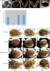
CTRP3/cartducin AS-ODN-treated organ culture system during the formation of condylar cartilage and Meckel's cartilage. (A) Procedures of tissue preparation. (B) Real-time RT-PCR for CTRP3/cartducin mRNA in condylar cartilage explants culture with 30 μm of AS-ODN. The level of CTRP3/cartducin mRNA expression in the AS-ODN group decreased on average by 97% that of the ODN-free group. Data are presented as means ± SD (n = 3). Asterisks indicate statistical significance by Tukey's comparison test (P ≤ 0.01). (C–N) Organ-cultured explants of ODN-free groups (C–E), with S-ODN (F–H), Random-ODN (I–K) and AS-ODN (L–N) at time 0 (C,F,I,L), cultured for 1 day (D,G,J,M) and 4 days (E,H, K,N). Meckel's cartilage in the AS-ODN group showed curvature deformation by day 4 (arrows in M and N). Condylar anlagen in the AS-ODN group showed a flattened, tongue-like appearance by day 4 (arrowheads in M and N). MC, Meckel's cartilage; CA, condylar anlagen. Bar: 500 μm.
Fig. 1
Fig. 1
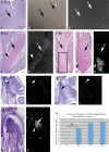
(A–H) Longitudinal sections of anlagen of Meckel's cartilage at E11.5 (A–C), formed Meckel's cartilage at E14.0 (D,E) and E16.0 (F–H) with toluidine blue staining (A,D,F,G) in situ hybridization of CTRP3/cartducin mRNA in bright field (B) and dark field (C,F,H). (G) Higher magnification of rectangular area in F. The anlagen of Meckel's cartilage did not express CTRP3/cartducin mRNA (arrows in B and C). CTRP3/cartducin mRNA was detected in clearly formed Meckel's cartilage (arrow in E), but not in the perichondrium (arrowheads in E and H). CTRP3/cartducin mRNA was reduced in the hypertrophic chondrocytes (arrow in H). Bar: 100 μm. (I–N) Coronal section of condylar anlagen at E14.0 (I,J), and formed condylar cartilage at E15.0 (K,L) and E16. 0 (M,N). Toluidine blue staining (I, K,M) and in situ hybridization of CTRP3/cartducin mRNA in dark field (J, L, N). CTRP3/cartducin mRNA was not detected in the condylar anlagen (arrowhead in J) but was expressed in the embryonic zone and in the upper part of condylar cartilage (arrowhead in L), and reduced in the lower part of hypertrophic cell zone (asterisk in N). F, fibrous cell zone; P, polymorphic cell zone; Fl, flattened cell zone; H, hypertrophic cell zone. Bar: 100 μm. (O) Real-time RT-PCR for CTRP3/cartducin mRNA in condylar anlagen/cartilage. Little CTRP3/cartducin mRNA expression was recognized in the condylar anlagen at E14.0. CTRP3/cartducin mRNA expression levels were significantly upregulated at E16.0, following which they showed a more gradual increase up to postnatal day 1. Data are presented as means ± SD (n = 3). Asterisks indicate statistical significance by Tukey's comparison test (P ≤ 0.01).
Fig. 3
Fig. 3
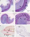
General histology of Meckel's cartilage of ODN-free controls (A,B,E) and AS-ODN-treated explants (C,D,F) after 4-day culture with toluidine blue staining in Technovit sections (A–D) and Van Gieson staining in paraffin sections (E,F). (B and D) Higher magnification of rectangular area in A and C, respectively. Inset in D is higher magnification of rectangular area in D. Well organized perichondrium was observed in the control (arrows in B). Loss of perichondrium and a newly formed, metachromatically stained matrix was seen at the position where the perichondrium had existed in the AS-ODN group (arrows in D). Cells within the metachromatically stained matrix were large and resembled chondrocytes (inset in D). Muscle tissue (arrowheads in E) and osteoid-like tissue (arrow in E) were formed around Meckel's cartilage (MC) in the control, but not in the AS-ODN group (F). Bars: 200 μm (A,E), 100 μm (B), 10 μm (inset in D).
Fig. 4
Fig. 4
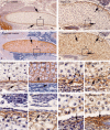
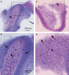
Immunohistochemistry for type I collagen (A,B,E,G,I,K) and for aggrecan (C,D,F,H,J,L) in Meckel's cartilage in ODN-free controls (A,C,E,F) and AS-ODN-treated explants (B,D,G–L) after 4-day culture. (E–J) Higher magnification of rectangular areas in A–D, respectively; K and L are from another sample. Co-localization of type I collagen and aggrecan immunostaining was recognized in the borderline between cartilage and perichondrium in the control (arrowheads in E and F). The borderline was lost and co-localization of type I collagen and aggrecan immunostaining was recognized in the intercellular matrix around chondrocyte-like cells (CLC) in the peripheral areas of AS-ODN-treated Meckel's cartilage (arrows in G–J). Co-localization of type I collagen and aggrecan immunostaining was occasionally observed in the remaining perichondrium (arrows in K and L). Bar: 100 μm.
Fig. 5
Fig. 5
General histology of condylar cartilage of ODN-free controls (A,B) and AS-ODN-treated explants (C,D) after 4-day culture with toluidine blue staining in Technovit sections. (B,D) Higher magnification of rectangular area in (A,C), respectively. Well-organized condylar cartilage (arrowhead in B) with perichondrium (arrows in B) was formed in the control group. Distinct architecture of zones was recognized. AS-ODN-treated condylar cartilage lacked the perichondrium and the fibrous cell zone (arrows in D). A distinct architecture of zones was lost (arrowhead in D). F, fibrous cell zone; P, polymorphic cell zone; Fl, flattened cell zone; H, hypertrophic cell zone. Bars: 200 μm (A,B) and 100 μm (C,D).
Fig. 6
Fig. 6
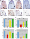
Immunostaining for aggrecan (A,E), tenascin-C (B,F), type I collagen (C,G), and type X collagen (D,H) in condylar cartilage of ODN-free controls (A–D) and AS-ODN-treated explants (E–H) after 4-day culture, and real-time RT-PCR analysis of mRNA for each cartilage matrix protein (I–L) in AS-ODN group and three controls including the ODN-free, S-ODN and Random-ODN groups. (A–H) Each immunostaining in control condylar cartilage was detected in restricted zones as recognized in vivo, but immunostaining for aggrecan, tenascin-C, and type I collagen was detected throughout the cartilage matrix, and immunostaining for type X collagen was not detected in AS-ODN-treated condylar cartilage. F, fibrous cell zone; P, polymorphic cell zone; Fl, flatted cell zone; H, hypertrophic cell zone. Bar: 100 μm. (I–L) The expression level of aggrecan, tenascin-C, collagen types I and X mRNA in the AS-ODN-treated condylar cartilage significantly decreased compared with ODN-free condylar cartilage (I–L). Data are presented as means ± SD (N = 3). Asterisks indicate statistical significance by Tukey's comparison test (*P < 0.05, **P < 0.01).
Fig. 7
Fig. 7
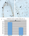
Cell proliferation assay in organ-cultured condylar cartilage. BrdU labeling in the chondrocytes of ODN-free control (A) and AS-ODN-treated explants (B). BrdU-positive cells were diffusely distributed throughout the entire cartilaginous tissue (arrowheads in B). The ratio of BrdU-positive cell number to total cells in AS-ODN-treated condylar cartilage number from whole sections was significantly reduced to 59.5% of that in ODN-free condylar cartilage (C). F, fibrous cell zone; P, polymorphic cell zone; Fl, flatted cell zone; H, hypertrophic cell zone. Data are presented as means ± SD (n = 4). Asterisks indicate statistical significance by Student's t-test (*P < 0.05). Bar: 100 μm.
Fig. 8
Fig. 8
Ultrastructure of cartilage matrix of control (A,C) and AS-ODN-treated explants (B,D) in Meckel's cartilage (A,B) and condylar cartilage (C,D). Density of thin collagen fibrils (arrows in A and B) and fine proteoglycan granules (arrowheads in A and B) was decreased in Meckel's cartilage in AS-ODN group (compare A with B). Density of relatively thick collagen fibrils (arrows in C and D) and proteoglycan granules (arrowheads in C and D) was remarkably decreased in condylar cartilage of AS-ODN-treated explants (compare C with D). Bar: 1 μm.
Fig. 9
Fig. 9
Organ-cultured explants (A–F) and their general histology (toluidine blue staining) of Meckel's cartilage (G,H, K, L) and condylar cartilage (I,J,M,N) with recombinant CTRP/cartducin protein with AS-ODN (A,B,G,I), without ODN (ODN-free) (D,E, K,M) on day 1(A,D) and on day 4 (B,E,G,I,K,M). Organ-cultured explants with just AS-ODN (C,H,J) and without ODN (ODN-free) (F,L,N) on day 4. Reduced curvature deformation in Meckel's cartilage (arrow in B) and remaining perichondrium of Meckel's cartilage (arrow in G) and condylar cartilage (arrows in I) were observed in recombinant protein-treated explants with AS-ODN. No specific histological changes were observed in either type of cartilage in recombinant protein-treated explants (K,M) compared with ODN-free explants (L,N). CA, condylar anlagen; MC, Meckel's cartilage; CC, condylar cartilage. Bar: 100 μm.

Similar articles

Cited by

References

    1. Akiyama H, Furukawa S, Wakisaka S, et al. Cartducin stimulates mesenchymal chondroprogenitor cell proliferation through both extracellular signal-regulated kinase and phosphatidylinositol 3-kinase/Akt pathways. FEBS J. 2006;273:2257–2263. - PubMed
    1. Akiyama H, Furukawa S, Wakisaka S, et al. Elevated expression of CTRP3/cartducin contribute to promotion of osteosarcoma cell proliferation. Oncol Rep. 2009;21:1477–1481. - PubMed
    1. Beresford WA. Secondary cartilages I, II. In: Beresford WA, editor. Chondroid Bone, Secondary Cartilage and Metaplasia. Baltimore: Urban and Schwarzenberg; 1981. pp. 23–65.
    1. Fukada K, Shibata S, Suzuki S, et al. In situ hybridization study of type I, II, X collagens and aggrecan mRNAs in the developing condylar cartilage of the fetal mouse mandible. J Anat. 1999;195:321–329. - PMC - PubMed
    1. Fukuoka H, Shibata S, Suda N, et al. Bone morphogenetic protein rescues lack of secondary cartilage in Runx2-deficient mice. J Anat. 2007;211:8–15. - PMC - PubMed

Publication types

LinkOut - more resources