

Review
doi: 10.1186/ar3206.
Disturbances of apoptotic cell clearance in systemic lupus erythematosus
Affiliations
- PMID: 21371352
- PMCID: PMC3157636
- DOI: 10.1186/ar3206
Item in Clipboard
Review
Disturbances of apoptotic cell clearance in systemic lupus erythematosus
Arthritis Res Ther.
.
Abstract
Systemic lupus erythematosus is a multifactorial autoimmune disease with an as yet unknown etiopathogenesis. It is widely thought that self-immunization in systemic lupus is driven by defective clearance of dead and dying cells. In lupus patients, large numbers of apoptotic cells accumulate in various tissues including germinal centers. In the present review, we discuss the danger signals released by apoptotic cells, their triggering of inflammatory responses, and the breakdown of B-cell tolerance. We also review the pathogenic role of apoptotic cell clearance in systemic lupus erythematosus.
Figures
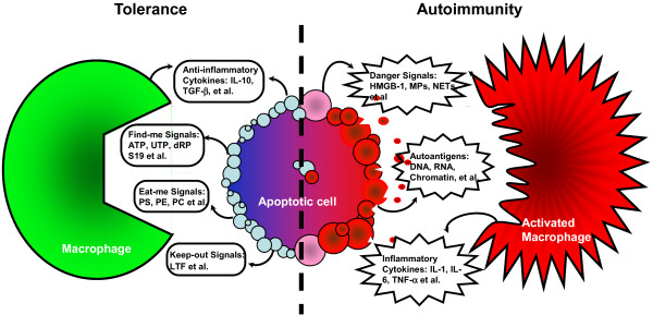
Distinct balance of apoptotic cell clearance. Normal clearance of apoptotic cells (left side, blue) involves sequential signals and plays an important role in tolerance induction and maintenance. Inflammatory clearance of apoptotic cells (right side, red) involves multi-inflammatory stimuli, breaks down tolerance, and drives autoimmunity including systemic lupus erythematosus. Blue blebs: early apoptotic cells modify surface markers and release signals to regulate chemotaxis and phagocytosis. Red blebs: later apoptotic cells and necrotic cells lose the cell membrane integrity, leading to the release of danger signals and modified autoantigens. dRP S19, dimer of ribosomal protein S19; HMGB1, high mobility group box 1; IL, interleukin; LTF, lactoferrin; MP, microparticle; NET, neutrophil extracellular trap; PC, phosphatidylcholine; PE, phosphatidylethanolamine; PS, phosphatidylserine; TGF-β, transforming growth factor beta.
References
-
- Horino K, Nishiura H, Ohsako T, Shibuya Y, Hiraoka T, Kitamura N, Yamamoto T. A monocyte chemotactic factor, S19 ribosomal protein dimer, in phagocytic clearance of apoptotic cells. Lab Invest. 1998;78:603–617. - PubMed
-
- Elliott MR, Chekeni FB, Trampont PC, Lazarowski ER, Kadl A, Walk SF, Park D, Woodson RI, Ostankovich M, Sharma P, Lysiak JJ, Harden TK, Leitinger N, Ravichandran KS. Nucleotides released by apoptotic cells act as a find-me signal to promote phagocytic clearance. Nature. 2009;461:282–286. doi: 10.1038/nature08296. - DOI - PMC - PubMed
Publication types
MeSH terms
Grants and funding
LinkOut - more resources
Full Text Sources
Medical
