. 2011 Mar 25;406(4):643-8.
doi: 10.1016/j.bbrc.2011.02.123. Epub 2011 Mar 1.

Elevated invasive potential of glioblastoma stem cells

Affiliations

Elevated invasive potential of glioblastoma stem cells

Lin Cheng et al. Biochem Biophys Res Commun. .

Abstract

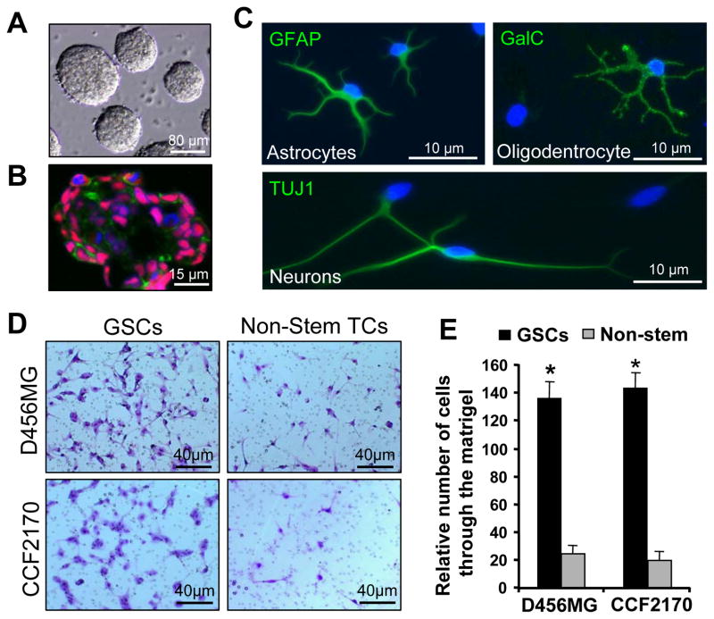
Glioblastomas (GBMs) are the most lethal and common types of primary brain tumors. The hallmark of GBMs is their highly infiltrative nature. The cellular and molecular mechanisms underlying the aggressive cancer invasion in GBMs are poorly understood. GBM displays remarkable cellular heterogeneity and hierarchy containing self-renewing glioblastoma stem cells (GSCs). Whether GSCs are more invasive than non-stem tumor cells and contribute to the invasive phenotype in GBMs has not been determined. Here we provide experimental evidence supporting that GSCs derived from GBM surgical specimens or xenografts display greater invasive potential in vitro and in vivo than matched non-stem tumor cells. Furthermore, we identified several invasion-associated proteins that were differentially expressed in GSCs relative to non-stem tumor cells. One of such proteins is L1CAM, a cell surface molecule shown to be critical to maintain GSC tumorigenic potential in our previous study. Immunohistochemical staining showed that L1CAM is highly expressed in a population of cancer cells in the invasive fronts of primary GBMs. Collectively, these data demonstrate the invasive nature of GSCs, suggesting that disrupting GSCs through a specific target such as L1CAM may reduce GBM cancer invasion and tumor recurrence.


Figures

Fig. 1
Fig. 1
Glioblastoma stem cells (GSCs) are invasive than matched non-stem tumor cells in vitro. (A) Representative images of GSC tumorspheres. GSCs derived from a primary GBM specimen (CCF2170) were cultured in neurobasal stem cell medium for seven days to form tumorspheres. (B) Immunofluorescent staining of SOX2 (a stem cell transcription factor) and L1CAM (a surface molecule of GSC) on frozen sections of GSC neurospheres. SOX2 was labeled in red, L1CAM in green, and nuclei were counterstained with DAPI in blue. (C) Immunofluorescent staining of GFAP (astrocyte marker), Galc (oligodentrocyte marker) or TUJ1 (neuronal marker) in the differentiated cells derived from GSCs. Isolated GSCs from a primary GBM (CCF2170) were induced for differentiation for 7 days and then immunostained with antibodies against GFAP, Galc or TUJ1 (green). Nuclei were counterstained with DAPI (blue). (D) In vitro matrigel invasion assay of GSCs and matched non-stem tumor cells from two GBMs. The relative invasive capacity of GSCs and matched non-stem tumor cells (Non-stem TCs) derived from D456MG GBM xenograft and CCF2170 primary GBM were examined in the BD Matrigel gel. Cells migrated through the matrigel were stained and photographed. (E) Quantified data from (D) shows that GSCs had significantly more cells migrated through the matrigel than matched non-stem tumor cells in vitro. Data are means ± SD (n=3). *, p<0.001.
Fig. 2
Fig. 2
GSCs displayed greater invasive capacity than matched non-stem tumor cells in vivo. (A and B) Aggressive cancer invasion into brain tissues at early stage (day 5) after GSC transplantation into mouse brains. GSCs and non-stem tumor cells (Non-stem TCs) derived from a GBM xenograft (D456MG) were transplanted into mouse brains trough intracranial injection (10,000 cells/mouse). Mouse brains harvested 5 days after tumor cell transplantation were examined for cancer cell dispersal through histological analysis. Aggressive cancer cell invasion (indicated by red arrows) into brain tissues were detected in mouse brains implanted with GSCs. But none cancer cell dispersal was found in the brains implanted with non-stem TCs, and all cells stayed in the injection site (indicated by a black arrow). (C and D) Infiltration of cancer cells into normal tissues in mouse brains implanted with GSCs at late stage (day 35) after cell transplantation. GSCs and non-stem tumor cells (Non-stem TCs) isolated from a D456MG GBM xenograft were transplanted into mouse brains trough intracranial injection (200,000 cells/mouse). Mouse brains were harvested 35 days after cell transplantation and then examined for cancer cell invasion through histological analysis. Diffuse infiltration of cancer cells (indicated by red arrows) into normal tissues were detected in brains implanted with GSCs but not in those with non-stem tumor cells, although D456MG non-stem tumors also grew small tumors (indicated by a black arrow) in mouse brains. (E and F) Representative images of brain cross sections (H & E stained) from mouse brains harvested on day 39 after cell transplantation described in (C and D). GSCs formed much larger tumors than matched non-stem tumor cells in mouse brains. Red arrows indicate large infiltrative tumors in the brain implanted with GSCs, and a black arrow indicates a small tumor in the brain implanted with non-stem tumor cells.
Fig. 3
Fig. 3
Immunoblot analysis shows increased expression of several invasion-related proteins in GSCs relative to matched non-stem tumor cells. GSCs and matched non-stem tumor cells (Non-stem TCs) isolated from a primary GBM (CCF1992) and a GBM xenograft (T3359) were analyzed for the expression of invasion-associated proteins including L1CAM, MMP16, ADAMTS1 and SEMA3C, and the GSC marker SOX2. The sizes of molecular markers are indicated on right side.
Fig. 4
Fig. 4
Immunohistochemical (IHC) staining shows that L1CAM is highly expressed in a subpopulation of cancer cells in the invasive fronts of primary GBMs. Representative images of L1CAM staining (in brown) in two GBMs (CCF1863 and T4121) are shown. The marked invasive fronts with small squares were enlarged in right panels to show L1CAM expression in the invasive fronts (indicated by arrows). The tumor sections were counterstained with hematoxylin. T, tumor tissue, N, normal brain tissue.

References

    1. Furnari FB, Fenton T, Bachoo RM, Mukasa A, Stommel JM, Stegh A, Hahn WC, Ligon KL, Louis DN, Brennan C, Chin L, DePinho RA, Cavenee WK. Malignant astrocytic glioma: Genetics, biology, and paths to treatment. Genes and Development. 2007;21:2683–2710. - PubMed
    1. Wen PY, Kesari S. Malignant gliomas in adults. New England Journal of Medicine. 2008;359:492–507. - PubMed
    1. Stupp R, Mason WP, Van Den Bent MJ, Weller M, Fisher B, Taphoorn MJB, Belanger K, Brandes AA, Marosi C, Bogdahn U, Curschmann J, Janzer RC, Ludwin SK, Gorlia T, Allgeier A, Lacombe D, Cairncross JG, Eisenhauer E, Mirimanoff RO. Radiotherapy plus concomitant and adjuvant temozolomide for glioblastoma. New England Journal of Medicine. 2005;352:987–996. - PubMed
    1. Park DM, Sathornsumetee S, Rich JN. Medical oncology: Treatment and management of malignant gliomas. Nature Reviews Clinical Oncology. 2010;7:75–77. - PubMed
    1. Hemmati HD, Nakano I, Lazareff JA, Masterman-Smith M, Geschwind DH, Bronner-Fraser M, Kornblum HI. Cancerous stem cells can arise from pediatric brain tumors. Proceedings of the National Academy of Sciences of the United States of America. 2003;100:15178–15183. - PMC - PubMed

Publication types

Substances