Review
. 2011 Aug;76(9):903-9.
doi: 10.1016/j.steroids.2011.02.011. Epub 2011 Mar 1.

Progesterone signaling mediated through progesterone receptor membrane component-1 in ovarian cells with special emphasis on ovarian cancer

Affiliations
Review

Progesterone signaling mediated through progesterone receptor membrane component-1 in ovarian cells with special emphasis on ovarian cancer

John J Peluso. Steroids. 2011 Aug.

Abstract

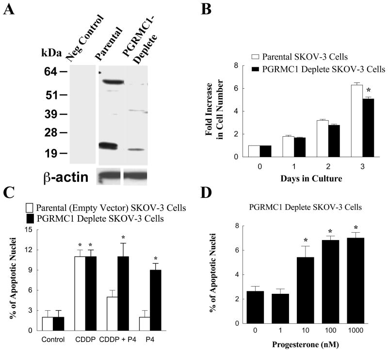
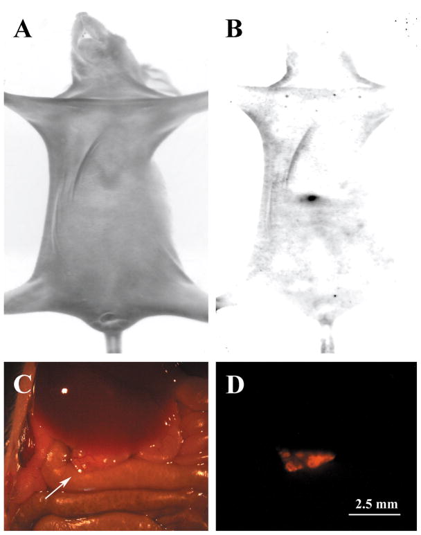
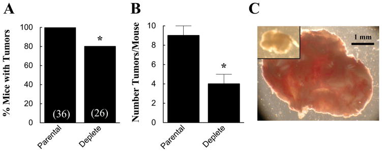
Various ovarian cell types including granulosa cells and ovarian surface epithelial cells express the progesterone (P4) binding protein, progesterone receptor membrane component-1 (PGRMC1). PGRMC1 is also expressed in ovarian tumors. PGRMC1 plays an essential role in promoting the survival of both normal and cancerous ovarian cell in vitro. Given the clinical significance of factors that regulate the viability of ovarian cancer, this review will focus on the role of PGRMC1 in ovarian cancer, while drawing insights into the mechanism of PGRMC1's action from cell lines derived from healthy ovaries as well as ovarian tumors. Studies using PGRMC1siRNA demonstrated that P4's ability to inhibit ovarian cells from undergoing apoptosis in vitro is dependent on PGRMC1. To confirm the importance of PGRMC1, the ability of PGRMC1-deplete ovarian cancer cell lines to form tumors in intact nude mice was assessed. Compared to PGRMC1-expressing ovarian cancer cells, PGRMC1-deplete ovarian cancer cells formed tumors in fewer mice (80% compared to 100% for controls). Moreover, the number of tumors derived from PGRMC1-deplete ovarian cancer cells was 50% of that observed in controls. Finally, the tumors that formed from PGRMC1-deplete ovarian cancer cells were about a fourth the size of tumors derived from ovarian cancer cells with normal levels of PGRMC1. One reason for PGRMC1-deplete tumors being smaller is that they had a poorly developed microvasculature system. How PGRMC1 regulates cell viability and in turn tumor growth is not known but part of the mechanism likely involves the regulation of genes that promote cell survival and inhibit apoptosis.


Conflict of interest statement

Disclosure Statement

The author does not have any actual or potential conflicts of interest.

Figures

Figure 1
Figure 1
Immunohistochemical localization of Progesterone Receptor (PGR) (A,B) and Progesterone Receptor Membrane Component-1 (PGRMC1) (C,D) in Stage IIIc Grade 2 (A,C) and Stage IIIc Grade 3 (B,D,E) ovarian tumors. Images from panels A and C and B and D are taken from adjacent sections from respective tumors. Panels A, B C and D are shown at the same magnification. The arrows in panels A and C mark the location of ovarian cancer cells that do not express PGR but do express PGRMC1. The image in panel E is a higher magnification of the image shown in panel D. Panel F is a negative control. Both panel E and F are shown at the same magnification. Data taken from Peluso et al [20].
Figure 2
Figure 2
The development of a dsRed-SKOV-3 cell line that was depleted in PGRMC1. Panel A is a western blot showing PGRMC1 levels in parental and PGRMC1-deplete dsRed SKOV-3 cells. β-actin western blot is shown to demonstrate equal protein loading. The − sign indicates a lane of a western blot in which the primary antibody was omitted and replaced with IgG (i.e. a negative control). The rate of cell proliferation of parental and PGRMC1-deplete SKOV-3 cells is shown in panel B. The effect of cisplatin (CDDP; 30 nM) and progesterone (P4; 1 μM) on the rate of apoptosis of parental empty vector and PGRMC1-deplete SKOV-3 cells is shown in panel C. The effect of increasing concentrations of P4 on the percentage of PGRMC1-deplete SKOV-3 cells undergoing apoptosis is shown in panel D. Data taken from Peluso et al [11].
Figure 3
Figure 3
Detection of ovarian tumors within intact living nude mice. The mouse shown in these images was injected with 10 million dsRed SKOV-3 cells and the tumors were allowed to develop for 5 weeks. In panel A, a bright field image of a nude mouse is shown. When observed under dsRed fluorescent filters (B), a tumor was observed (black arrow). This animal was dissected to expose the peritoneum and a tumor (arrow) was observed under bright field (C) and fluorescent optics (D).
Figure 4
Figure 4
The effect of depleting PGRMC1 on the percentage of mice that develop tumors (A) and the number of tumors, which develop in mice that form tumors (B). The numbers in parentheses in A are the number of mice observed. The effect of depleting PGRMC1 on the relative size of the tumors is shown in panel C. The tumor derived from PGRMC1-deplete SKOV-3 cells is show in the inset of panel C. Both images are shown at the same magnification. The data in panel A and B is taken from Peluso et al [11].
Figure 5
Figure 5
The effects of PGRMC1 siRNA treatment of the expression profile of various apoptosis-related genes is shown in panel A. The effects of 1 μM P4 plus scramble siRNA or 1 μM P4 plus PGRMC1 siRNA treatment are shown in panels B and C, respectively. In each graph the mRNA levels of apoptosis-related genes were assessed by real-time PCR and shown as a percentage change from scramble control values. The values are means (± standard error) of three experiments. Genes associated with the induction of apoptosis are shown in red and the anti-apoptotic genes shown in green. Data is taken from Peluso et al [25].

Similar articles

Cited by

References

    1. Salzberg M, Thurlimann B, Bonnefois H, Fink D, Rochlitz C, von Moos R, Senn H. Current concepts of treatment strategies in advanced or recurrent ovarian cancer. Oncology. 2005;68:293–298. - PubMed
    1. Ho SM. Estrogen, progesterone and epithelial ovarian cancer. Reprod Biol Endocrinol. 2003;1:73. - PMC - PubMed
    1. Fauvet R, Dufournet Etienne C, Poncelet C, Bringuier AF, Feldmann G, Darai E. Effects of progesterone and anti-progestin (mifepristone) treatment on proliferation and apoptosis of the human ovarian cancer cell line, OVCAR-3. Oncol Rep. 2006;15:743–748. - PubMed
    1. Syed V, Mukherjee K, Godoy-Tundidor S, Ho SM. Progesterone induces apoptosis in TRAIL-resistant ovarian cancer cells by circumventing c-FLIPL overexpression. J Cell Biochem. 2007;102:442–452. - PubMed
    1. Murdoch WJ, Van Kirk EA, Isaak DD, Shen Y. Progesterone facilitates cisplatin toxicity in epithelial ovarian cancer cells and xenografts. Gynecol Oncol. 2008;110:251–255. - PMC - PubMed

Publication types