. 2011 May;95(6):2087-93.
doi: 10.1016/j.fertnstert.2011.01.160. Epub 2011 Mar 3.

Temporal expression pattern of progesterone receptor in the uterine luminal epithelium suggests its requirement during early events of implantation

Affiliations

Temporal expression pattern of progesterone receptor in the uterine luminal epithelium suggests its requirement during early events of implantation

Honglu Diao et al. Fertil Steril. 2011 May.

Abstract

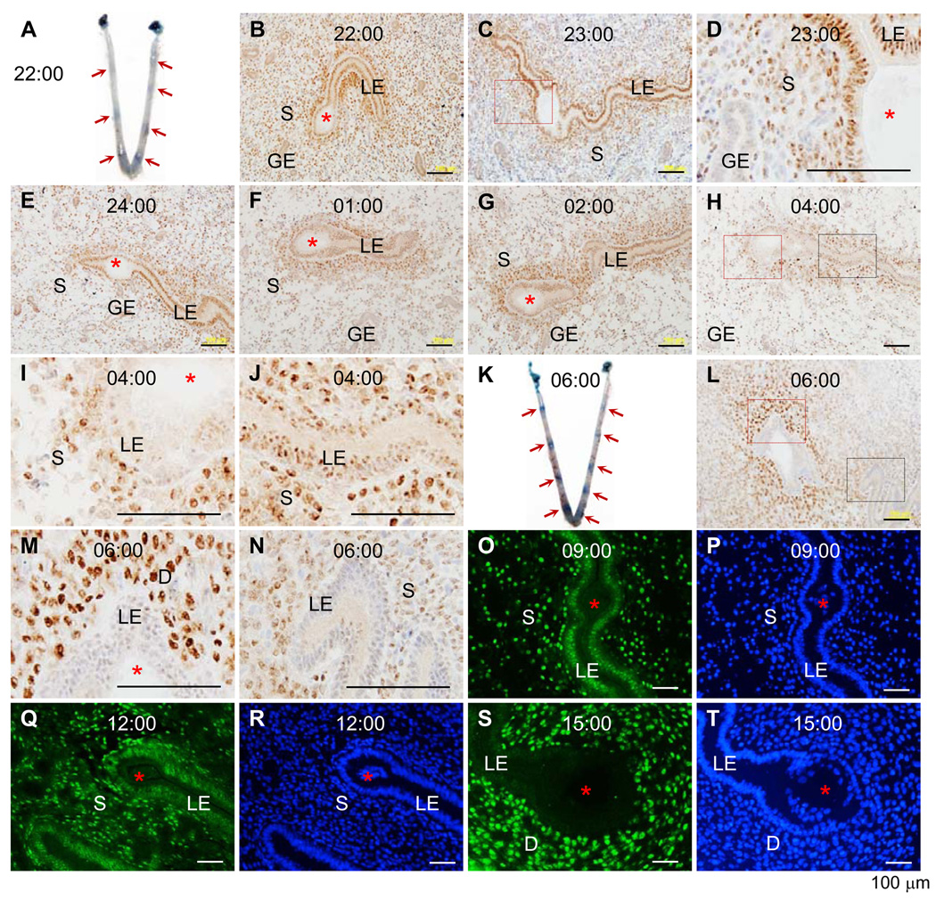
Objective: To determine the precise timing of progesterone receptor (PR) disappearing from the uterine luminal epithelium (LE) to help understand the significance of the dynamic PR expression in the LE during embryo implantation.

Design: Experimental rodent models.

Setting: University research laboratories.

Animal(s): Mice and hamsters.

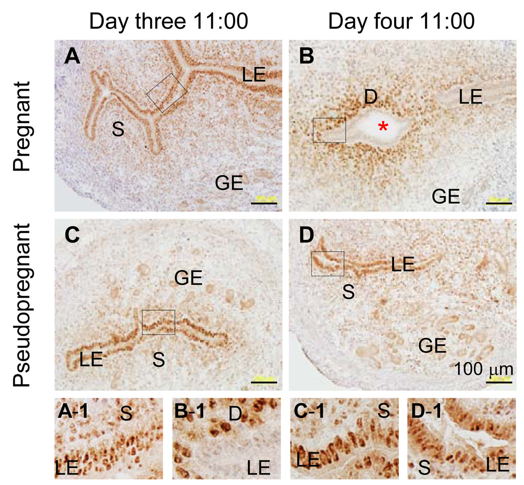
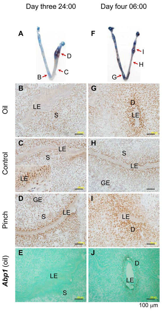
Intervention(s): Pseudopregnancy and artificial decidualization.

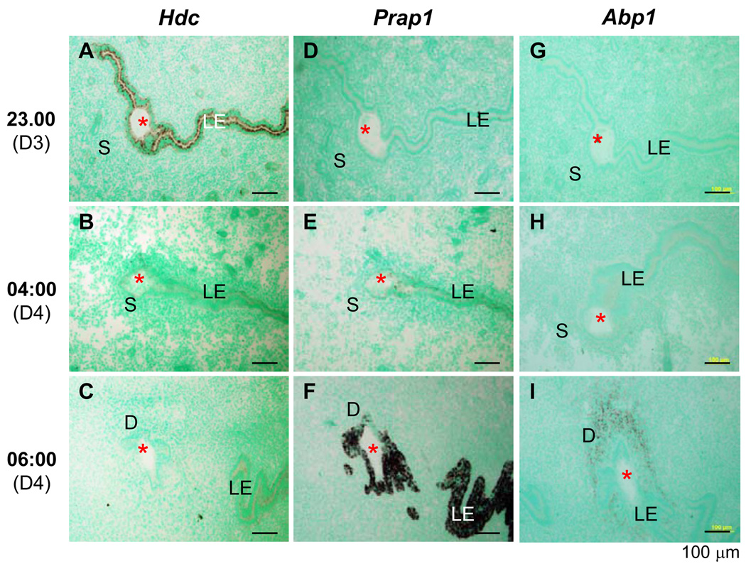
Main outcome measure(s): Blue dye injection for detecting embryo attachment; immunohistochemistry, immunofluorescence, and in situ hybridization for detecting gene expression.

Result(s): Progesterone receptor remained expressed in the LE up to 6 hours after the initial detection of blue dye reaction in mice (day 3, 22:00 hours), but disappeared first from LE cells at the implantation site and subsequently from the entire LE layer by day 4, 06:00 hours, when uterine stromal decidualization had become obvious. Progesterone receptor remained highly expressed in the LE of day 4 at 11:00 hours in pseudopregnant mice, but it disappeared from the entire LE layer by day 4 at 06:00 hours in artificially decidualized pseudopregnant mice.

Conclusion(s): Progesterone receptor disappears from the LE after implantation has initiated and before the histologic decidualization manifests, suggesting an active role of continued PR expression in the LE for the initial implantation process. The disappearance of PR expression in the LE is regulated by uterine factor(s) produced upon embryo attachment.


Figures

Figure 1
Figure 1
Time-course expression of progesterone receptor (PR) during the early hours of embryo implantation in mouse (A~N) uterus detected by immunohistochemistry (IHC) and hamster (O~T) uterus detected by immunofluorescence (IF). Frozen sections (10 µm) through an embryo were used. A. A representative uterine image on day three 22:00 hours. B. PR IHC of an implantation site on A. C. PR IHC, day three 23:00 hours. D. An enlarged view of the red boxed area in C. E. PR IHC, day three 24:00 hours. F. PR IHC, day four 01:00 hour. G. PR IHC, day four 02:00 hours. H. PR IHC, day four 04:00 hours. I. An enlarged view of the red boxed area in H. J. An enlarged view of the black boxed area in H. K. A representative uterine image on day four 06:00 hours. L. PR IHC of an implantation site on K. M. An enlarged view of the red boxed area in L. N. An enlarged view of the black boxed area in L. O. PR IF, day three 09:00 hours. P. DAPI stain of the section on O. Q. PR IF, day three 12:00 hours. R. DAPI stain of the section on Q. S. PR IF, day three 15:00 hours. T. DAPI stain of the section on S. Red arrows indicate implantation sites detected by blue dye reaction (A & K). LE, luminal epithelium; S, stroma; GE, glandular epithelium; D, decidual zone; red asterisk, embryo; scale bar, 100 µm. N=3–4. No specific staining was detected in the negative control (data not shown).
Figure 2
Figure 2
Time-course expression of histidine decarboxylase (Hdc), proline-rich acidic protein 1 (Prap1), and amiloride-binding protein 1 (Abp1) during the early hours of embryo implantation in mouse uterus detected by in situ hybridization using antisense probes. Longitudinal frozen sections (10 µm) through an embryo were used. A. Hdc, day three 23:00 hours. B. Hdc, day four 04:00 hours. C. Hdc, day four 06:00 hours. D. Prap, day three 23:00 hours. E. Prap1, day four 04:00 hours. F. Prap1, day four 06:00 hours. G. Abp1, day three 23:00 hours. H. Abp1, day four 04:00 hours. I. Abp1, day four 06:00 hours. LE, luminal epithelium; S, stroma; D, decidual zone; red asterisk, embryo; scale bar, 100 µm. N=3. No specific staining was detected with sense probes (data not shown).
Figure 3
Figure 3
Immnuohistochemistry of progesterone receptor in the normal pregnant and pseudopregnant mouse uterus on day three 11:00 hours and day four 11:00 hours. An enlarged view of each of the boxed area in A–D is shown on the smaller panels A-1 to D-1, respectively. A & A-1. NP, day three 11:00 hours. B & B-1. NP, day four 11:00 hours. C & C-1. PP, day three 11:00 hours. D & D-1. PP, day four 11:00 hours. LE, luminal epithelium; S, stroma; GE, glandular epithelium; D, decidual zone; red asterisk, embryo; scale bar, 100 µm. N=3. No specific staining was detected in the negative control (data not shown).
Figure 4
Figure 4
Immnuostaining of progesterone receptor (PR) and in situ hybridization (ISH) detection of decidualization marker amiloride-binding protein 1 (Abp1) in mouse uterus with artificial decidualization. Longitudinal frozen sections (10 µm) crossing luminal epithelium were used. A. A representative uterine image on day three 24:00 hours: left uterine horn with oil injection, right uterine horn with two pinches near the uterotubal junction, red arrows indicating the areas sectioned for PR immunohistochemistry (IHC) in B~D. B. PR IHC, day three 24:00 hours, oil-injected uterine horn. C. PR IHC, day three 24:00 hours, control uterine segment without stimulation; an enlarged view of the white boxed area shown in the insert on left bottom corner; the seemingly multiple luminal epithelial (LE) layers indicating a particular sectioning angle that cut through several LE cells on the single LE layer. D. PR IHC, day three 24:00 hours, a pinched area. E. Abp1, ISH, antisense probe, day three 24:00 hours, the same oil injected area as in B. Abp1 is undetectable at this hour. F. A representative uterine image on day four 06:00 hours: left uterine horn with oil injection, right uterine horn with two pinches near the uterotubal junction, red arrows indicating the areas sectioned for PR IHC in G~I. G. PR IHC, day four 06:00 hours, oil-injected uterine horn. H. PR IHC, day four 06:00 hours, control uterine segment without stimulation. I. PR IHC, day four 6:00 hours, a pinched area. J. Abp1, ISH, antisense probe, day four 06:00 hours, the same oil injected area as in G. Abp1 is detectable in the subepithelial stromal area at this hour. LE, luminal epithelium; S, stroma; GE, glandular epithelium; D, decidual zone; scale bar, 100 µm. N=3. No specific staining was detected in the negative control for PR IHC or using a sense probe for Abp1 ISH (data not shown).

Similar articles

Cited by

References

    1. Tan J, Paria BC, Dey SK, Das SK. Differential uterine expression of estrogen and progesterone receptors correlates with uterine preparation for implantation and decidualization in the mouse. Endocrinology. 1999;140:5310–5321. - PMC - PubMed
    1. Cheon YP, Li Q, Xu X, DeMayo FJ, Bagchi IC, Bagchi MK. A genomic approach to identify novel progesterone receptor regulated pathways in the uterus during implantation. Mol Endocrinol. 2002;16:2853–2871. - PubMed
    1. Bazer FW, Slayden OD. Progesterone-induced gene expression in uterine epithelia: a myth perpetuated by conventional wisdom. Biol Reprod. 2008;79:1008–1009. - PubMed
    1. Wang H, Dey SK. Roadmap to embryo implantation: clues from mouse models. Nat Rev Genet. 2006;7:185–199. - PubMed
    1. Mulac-Jericevic B, Conneely OM. Reproductive tissue selective actions of progesterone receptors. Reproduction. 2004;128:139–146. - PubMed

Publication types

Substances