

Vascular pathology of medial arterial calcifications in NT5E deficiency: implications for the role of adenosine in pseudoxanthoma elasticum
- PMID: 21371928
- PMCID: PMC3081917
- DOI: 10.1016/j.ymgme.2011.01.018
Vascular pathology of medial arterial calcifications in NT5E deficiency: implications for the role of adenosine in pseudoxanthoma elasticum
Abstract
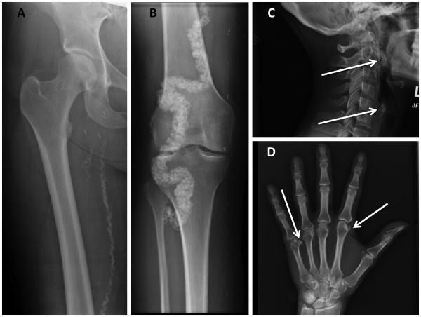
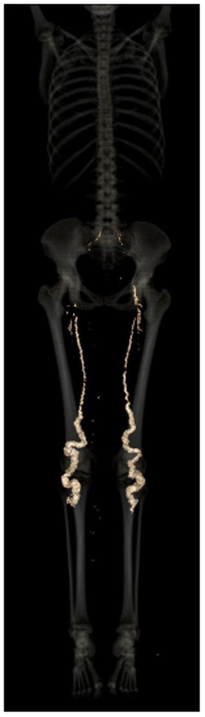
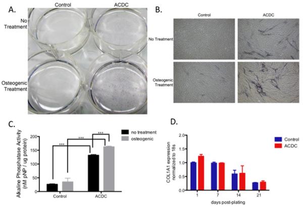
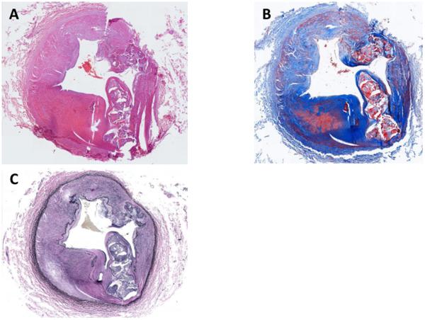
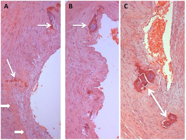
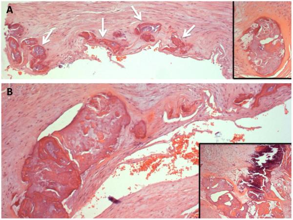
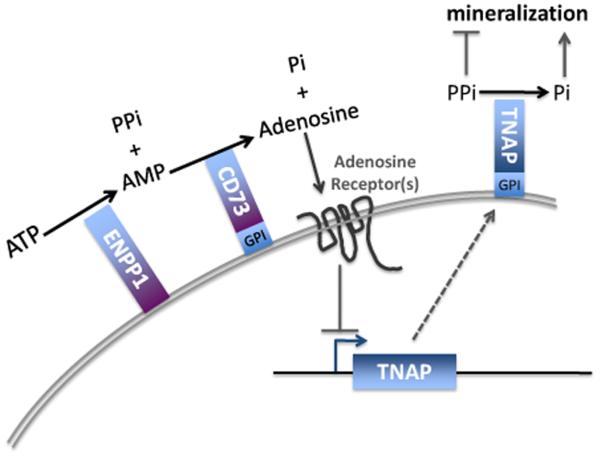
Arterial Calcification due to Deficiency of CD73 (ACDC) results from mutations in the NT5E gene encoding the 5' exonucleotidase, CD73. We now describe the third familial case of ACDC, including radiological and histopathological details of the arterial calcifications. The medial lesions involve the entire circumference of the elastic lamina, in contrast to the intimal plaque-like disease of atherosclerosis. The demonstration of broken and fragmented elastic fibers leading to generalized vascular calcification suggests an analogy to pseudoxanthoma elasticum (PXE), which exhibits similar histopathology. Classical PXE is caused by deficiency of ABCC6, a C type ABC transporter whose ligand is unknown. Other C type ABC proteins transport nucleotides, so the newly described role of adenosine in inhibiting vascular calcification, along with the similarity of ACDC and PXE with respect to vascular pathology, suggests that adenosine may be the ligand for ABCC6.
Published by Elsevier Inc.
Figures
Comment in
-
Reply to the article of C. Markello et al. entitled "Vascular pathology of medial arterial calcifications in NT5E deficiency: Implications for the role of adenosine in pseudoxanthoma elasticum".Mol Genet Metab. 2011 Jun;103(2):199-200. doi: 10.1016/j.ymgme.2011.03.014. Epub 2011 Mar 24. Mol Genet Metab. 2011. PMID: 21497121 Free PMC article. No abstract available.
References
-
- Von Magnus-Levy A. Ueber ungewöhnliche Verkalkung der Arterien. Deutsche Meidzinische Wochenschrift. 1914;40:1305–1309.
-
- Hilaire C, Ziegler SG, Markello T, Brusco A, Groden C, Gill F, Carlson-Donohoe H, Lederman RJ, Chen MY, Yang D, Siegenthaler MP, Arduino C, Mancini C, Freudenthal B, Stanescu HC, Zdebik AA, Chaganti RK, Nussbaum R, Kleta R, Gahl WA, Boehm M. NT5E mutations and arterial calcifications. N. Engl. J. Med. 2010 in press. - PMC - PubMed
-
- Yegutkin GG. Nucleotide- and nucleoside-converting ectoenzymes: Important modulators of purinergic signaling cascade. Biochim. Biophys. Acta. 2008;1783:673–694. - PubMed
-
- Truter S, Rosenbaum-Fiedler J, Sapadin A, Lebwohl M. Calcification of elastic fibers in pseudoxanthoma elasticum. Mt. Sinai J Med. 1996;63:210–215. - PubMed
Publication types
MeSH terms
Substances
Grants and funding
LinkOut - more resources
Full Text Sources
Other Literature Sources
Medical
Research Materials
Miscellaneous
