. 2011 Apr;134(Pt 4):1127-39.
doi: 10.1093/brain/awr025. Epub 2011 Mar 2.

JAK-STAT3 pathway regulates spinal astrocyte proliferation and neuropathic pain maintenance in rats

Affiliations

JAK-STAT3 pathway regulates spinal astrocyte proliferation and neuropathic pain maintenance in rats

Makoto Tsuda et al. Brain. 2011 Apr.

Abstract

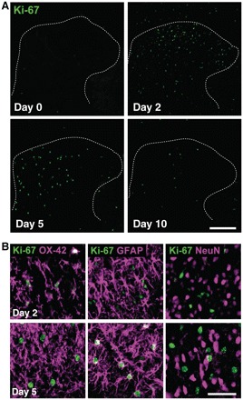
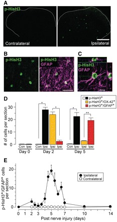
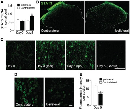
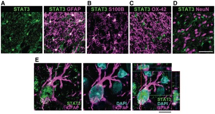
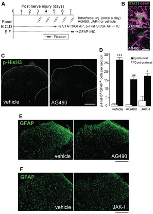
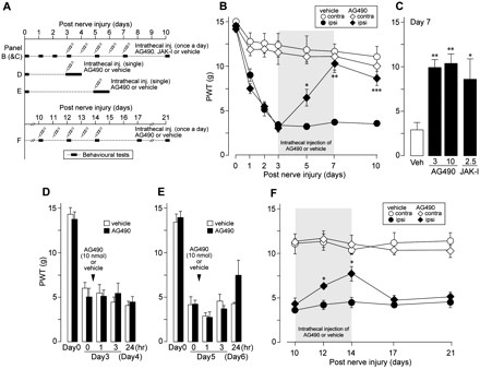
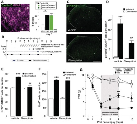
Neuropathic pain, a debilitating pain condition, is a common consequence of damage to the nervous system. Optimal treatment of neuropathic pain is a major clinical challenge because the underlying mechanisms remain unclear and currently available treatments are frequently ineffective. Emerging lines of evidence indicate that peripheral nerve injury converts resting spinal cord glia into reactive cells that are required for the development and maintenance of neuropathic pain. However, the mechanisms underlying reactive astrogliosis after nerve injury are largely unknown. In the present study, we investigated cell proliferation, a critical process in reactive astrogliosis, and determined the temporally restricted proliferation of dorsal horn astrocytes in rats with spinal nerve injury, a well-known model of neuropathic pain. We found that nerve injury-induced astrocyte proliferation requires the Janus kinase-signal transducers and activators of transcription 3 signalling pathway. Nerve injury induced a marked signal transducers and activators of transcription 3 nuclear translocation, a primary index of signal transducers and activators of transcription 3 activation, in dorsal horn astrocytes. Intrathecally administering inhibitors of Janus kinase-signal transducers and activators of transcription 3 signalling to rats with nerve injury reduced the number of proliferating dorsal horn astrocytes and produced a recovery from established tactile allodynia, a cardinal symptom of neuropathic pain that is characterized by pain hypersensitivity evoked by innocuous stimuli. Moreover, recovery from tactile allodynia was also produced by direct suppression of dividing astrocytes by intrathecal administration of the cell cycle inhibitor flavopiridol to nerve-injured rats. Together, these results imply that the Janus kinase-signal transducers and activators of transcription 3 signalling pathway are critical transducers of astrocyte proliferation and maintenance of tactile allodynia and may be a therapeutic target for neuropathic pain.


Figures

Figure 1
Figure 1
Immunohistochemical characterization of proliferating cells in the dorsal horn after peripheral nerve injury in rats. (A) Immunofluorescence of Ki-67, a nuclear protein expressed in all cell cycle phases except the resting phase, in fifth lumbar dorsal horn sections on Day 0, 2, 5 and 10 after peripheral nerve injury. (B) Double immunofluorescence labelling for Ki-67 (green) and cell-type markers (magenta: OX-42, microglia; GFAP, astrocytes; neuronal nuclei, neurons) in fifth lumbar dorsal horn sections on Day 2 (upper three panels) and Day 5 (lower three panels) post-peripheral nerve injury. Scale bar = 200 μm (A), 50 μm (B).
Figure 2
Figure 2
Mitotic phase of cycling astrocytes in the dorsal horn after peripheral nerve injury in rats. (A) Immunofluorescence of p-HisH3, a marker for G2/M phase of the cell cycle, in fifth lumbar dorsal horn sections 5 days after peripheral nerve injury. (B) Double immunofluorescence labelling for p-HisH3 (green) and GFAP (magenta) shown in a single channel (p-HisH3, left) and as a merged image (p-HisH3/GFAP, right) from grey matter of the fifth lumbar dorsal horn 5 days after peripheral nerve injury. (C) Representative confocal z-stack digital images of a single cell double-immunolabelled with p-HisH3 (green) and GFAP (magenta). (D) The numbers of p-HisH3+ cells (open and closed columns), p-HisH3+/OX-42+ cells (yellow columns) and p-HisH3+/GFAP+ cells (red columns) in fifth lumbar dorsal horn sections from rats 0, 2 and 5 days after peripheral nerve injury. Values represent the number of cells (per dorsal horn) (n = 4–6 rats; *P < 0.05, **P < 0.01). (E) The time course of p-HisH3+/GFAP+ cells in the dorsal horn after peripheral nerve injury. Values represent the number of p-HisH3+/GFAP+ cells (per dorsal horn) (n = 3–5 rats per each time point; *P < 0.05, **P < 0.01 versus contralateral side at the corresponding time point). Scale bar = 200 μm (A), 50 μm (B), 10 μm (C). Data are mean ± SEM.
Figure 3
Figure 3
STAT3 expression in the dorsal horn and dorsal root ganglion after peripheral nerve injury in rats. (A) Real-time quantitative polymerase chain reaction analysis of STAT3 messenger RNA in the total RNA extract from the rat spinal cord ipsilateral and contralateral to the peripheral nerve injury on Day 0 and 5 after peripheral nerve injury. The levels of STAT3 messenger RNA were normalized to the value of GAPDH messenger RNA, and values represent the ratio of STAT3 messenger RNA/GAPDH messenger RNA (n = 4 rats; *P < 0.05 versus contralateral side at the corresponding time point). (B) Immunofluorescence of STAT3 in fifth lumbar dorsal horn sections 5 days after peripheral nerve injury. (C) STAT3 immunofluorescence images at high-magnification from grey matter of the fifth lumbar dorsal horn 0, 3 and 5 days after peripheral nerve injury. (D) Immunofluorescence of STAT3 in fifth lumbar dorsal root ganglion sections 5 days after peripheral nerve injury. (E) Fluorescence intensity of STAT3 in the fifth lumbar dorsal root ganglion on Day 5 post-peripheral nerve injury (n = 3 rats; ***P < 0.001 versus contralateral side). Scale bar = 200 μm (B), 50 μm (C, D). Data are mean ± SEM.
Figure 4
Figure 4
STAT3 translocation to the nucleus of dorsal horn astrocytes after peripheral nerve injury in rats. (A–D) Double immunofluorescence labelling for STAT3 (green) and cell-type markers (magenta: A, GFAP; B, S100β; C, OX-42; D, neuronal nuclei) in fifth lumbar dorsal horn sections on Day 5 post-peripheral nerve injury. (E) Representative confocal z-stack digital images of a single cell triple labelled with STAT3 (green), GFAP (magenta) and DAPI (blue) shown in double channels (left, STAT3/GFAP; middle, DAPI/GFAP) and as a merged image (right, STAT3/GFAP/DAPI) from grey matter of the fifth lumbar dorsal horn 5 days after peripheral nerve injury. Scale bar = 50 μm (A–D), 10 μm (E).
Figure 5
Figure 5
Role of STAT3 signalling in dorsal horn astrocyte proliferation after peripheral nerve injury. (A) Schematic time-line for intrathecal administration and fixation. AG490 and JAK Inhibitor I, respective inhibitors of the STAT3 activator JAK, were administered intrathecally once a day for 2 days (B–D) and for 5 days (E and F) from Day 3 after peripheral nerve injury. (B) STAT3 immunofluorescence in representative images of GFAP+ cells in the fifth lumbar dorsal horn sections from vehicle- and AG490-treated rats. (C) p-HisH3 immunofluorescence in representative images of fifth lumbar dorsal horn sections from vehicle- and AG490-treated rats. (D) The numbers of p-HisH3+/GFAP+ cells in the fifth lumbar dorsal horn ipsilateral and contralateral to peripheral nerve injury from vehicle-, AG490- and JAK Inhibitor I-treated rats 5 days post-peripheral nerve injury. Values represent the number of p-HisH3+/GFAP+ cells (per dorsal horn) (n = 3–7 rats; ***P < 0.001 versus contralateral side of vehicle group; #P < 0.05, ##P < 0.01 versus ipsilateral side of vehicle group). (E and F) GFAP immunofluorescence in representative images of fifth lumbar dorsal horn sections from rats with peripheral nerve injury treated with either vehicle, AG490 or JAK Inhibitor I on postoperative Day 7. Scale bar = 10 μm (B), 200 μm (C, E and F). Data are mean ± SEM. IHC = immunohistochemistry.
Figure 6
Figure 6
Recovery from tactile allodynia after peripheral nerve injury by inhibiting JAK-STAT3 signalling in rats. (A) Schematic time-line for intrathecal administration and behavioural tests. (B) Rats with peripheral nerve injury were injected intrathecally with AG490 (10 nmol/10 μl) or vehicle (10 μl) once a day for 5 days from Day 3–7. Paw withdrawal threshold (PWT) to mechanical stimulation by von Frey filaments was measured before (Day 0), 1, 2, 3, 5, 7 and 10 days after peripheral nerve injury. Values represent the threshold (g) to elicit paw withdrawal behaviour (n = 5 rats; *P < 0.05, **P < 0.01, ***P < 0.001 versus vehicle group at corresponding time point). (C) Paw withdrawal threshold on Day 7 in rats with peripheral nerve injury-treated intrathecally with AG490 (3 or 10 nmol/10 μl), JAK Inhibitor I (2.5 nmol/10 μl) or vehicle (10 μl) once a day for 5 days from Day 3–7. Values represent the threshold (g) to elicit PWT (n = 5–7 rats; *P < 0.05, **P < 0.01 versus vehicle group). (D and E) Paw withdrawal threshold on Day 3 (D) and Day 5 (E) in rats with peripheral nerve injury with a single bolus intrathecal injection of AG490 (10 nmol/10 μl) or vehicle (10 μl). Paw withdrawal behaviour was measured before (Day 0), pre-injection (D, Day 3, 0 h; E, Day 5, 0 h), 1, 3 and 24 h after injection. Values represent the threshold (g) to elicit paw withdrawal behaviour (n = 4–6 rats). (F) Rats with peripheral nerve injury were injected intrathecally with AG490 (10 nmol/10 μl) or vehicle (10 μl) once a day for 5 days from Days 10–14. Paw withdrawal threshold was measured 10, 12, 14, 17 and 21 days after peripheral nerve injury. Values represent the threshold (g) to elicit paw withdrawal behaviour (n = 5 rats; *P < 0.05 versus vehicle group at corresponding time point). Data are mean ± SEM.
Figure 7
Figure 7
Effects of the cell cycle inhibitor flavopiridol on astrocyte proliferation and tactile allodynia after peripheral nerve injury. (A) Double immunofluorescence labelling for cyclin D1 (green) and GFAP (magenta) shown as a merged image from grey matter of the fifth lumbar dorsal horn 5 days after peripheral nerve injury. The numbers of cyclin D1+cells (open and closed columns), cyclin D1+/GFAP+ cells (green column) and cyclin D1+/GFAP cells (dark green column) in fifth lumbar dorsal horn sections from rats 5 days after peripheral nerve injury. Values represent the number of cells (per section) (n = 4 rats; *P < 0.05). (B) Schematic time-line for intrathecal administration, fixation and behavioural tests. (C) p-HisH3 immunofluorescence in representative images of fifth lumbar dorsal horn sections from vehicle- and flavopiridol-treated rats on Day 5 post-peripheral nerve injury. Rats with peripheral nerve injury were injected intrathecally with flavopiridol (5 nmol/10 μl) or vehicle (10 μl) twice a day for 2 days from Day 3. (D) The numbers of p-HisH3+/GFAP+ cells in the fifth lumbar dorsal horn ipsilateral and contralateral to peripheral nerve injury from vehicle- or flavopiridol-treated rats on 5 days post-peripheral nerve injury. Values represent the number of p-HisH3+/GFAP+ cells (per section) (n = 5 rats; ***P < 0.001 versus contralateral side of vehicle group; ##P < 0.01 versus ipsilateral side of vehicle group). (E and F) The numbers of GFAP+/S100β+ (E) and Iba1+ (F) cells in the fifth lumbar dorsal horn ipsilateral and contralateral to peripheral nerve injury from vehicle- or flavopiridol-treated rats on Day 7 post-peripheral nerve injury. Values represent the number of GFAP+/S100β+ (E) and Iba1+ (F) cells (per section) (n = 4 rats; ***P < 0.001 versus contralateral side of vehicle group; ##P < 0.01 versus ipsilateral side of vehicle group). (G) Paw withdrawal threshold (PWT) to mechanical stimulation by von Frey filaments was measured before (Day 0), and 1, 3, 5, 7 and 10 days after peripheral nerve injury. Rats with peripheral nerve injury were injected intrathecally with flavopiridol (5 nmol/10 μl) or vehicle (10 μl) twice a day for 5 days from Days 3 to 7. Values represent the threshold (g) to elicit paw withdrawal behaviour (n = 5 rats; *P < 0.05, **P < 0.01, ***P < 0.001 versus vehicle group at corresponding time point). Scale bar = 50 μm (A), 200 μm (C). Data are mean ± SEM. IHC = immunohistochemistry.

References

    1. Baron R. Mechanisms of disease: neuropathic pain–a clinical perspective. Nat Clin Pract Neurol. 2006;2:95–106. - PubMed
    1. Bonni A, Sun Y, Nadal-Vicens M, Bhatt A, Frank DA, Rozovsky I, et al. Regulation of gliogenesis in the central nervous system by the JAK-STAT signaling pathway. Science. 1997;278:477–83. - PubMed
    1. Byrnes KR, Stoica BA, Fricke S, Di Giovanni S, Faden AI. Cell cycle activation contributes to post-mitotic cell death and secondary damage after spinal cord injury. Brain. 2007;130:2977–92. - PubMed
    1. Chaplan SR, Bach FW, Pogrel JW, Chung JM, Yaksh TL. Quantitative assessment of tactile allodynia in the rat paw. J Neurosci Methods. 1994;53:55–63. - PubMed
    1. Choi JS, Kim SY, Cha JH, Choi YS, Sung KW, Oh ST, et al. Upregulation of gp130 and STAT3 activation in the rat hippocampus following transient forebrain ischemia. Glia. 2003;41:237–46. - PubMed

Publication types