

Characterization of soft contact lens edge fitting using ultra-high resolution and ultra-long scan depth optical coherence tomography
- PMID: 21372023
- PMCID: PMC3175978
- DOI: 10.1167/iovs.10-6507
Characterization of soft contact lens edge fitting using ultra-high resolution and ultra-long scan depth optical coherence tomography
Abstract
Purpose: To characterize the edge fitting of soft contact lenses using ultra-high resolution optical coherence tomography (UHR-OCT) and ultra-long scan depth optical coherence tomography (UL-OCT).
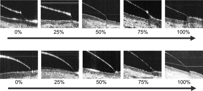
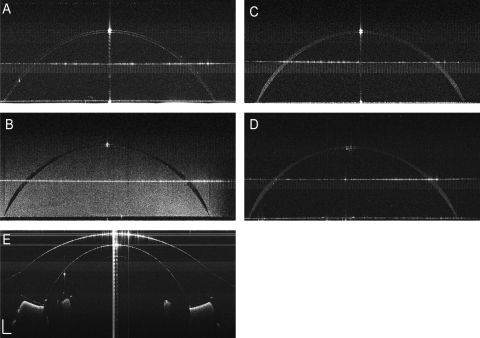
Methods: A total of 20 participants (11 men, 9 women; mean age, 32.3 years) were recruited. Four different types of soft contact lenses were randomly fitted to both eyes of each subject on two separate visits. After 30 minutes, the horizontal meridians of the corneal center, midperiphery, and limbus were imaged by UHR-OCT. UL-OCT imaged each lens in vitro and the ocular surface of a physical model eye.
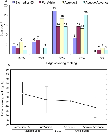
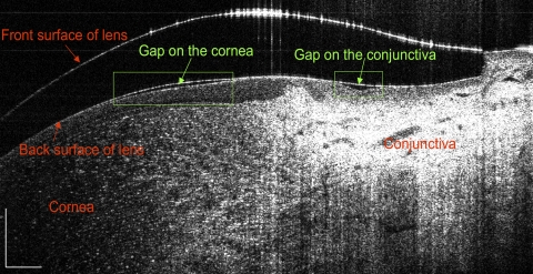
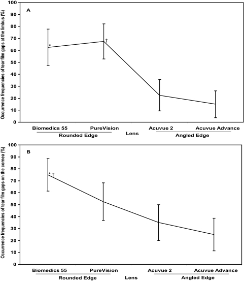
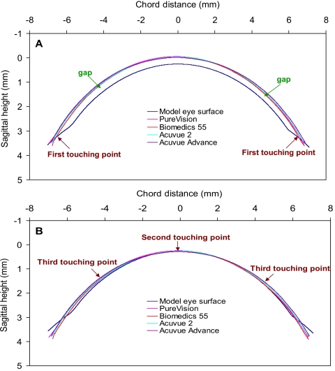
Results: Angle-edged lenses had significantly less conjunctival buildup than did round-edged lenses (P = 0.008). Limbal post-lens tear film gaps were present in 42% of the eyes, with the round-edged lenses having the most at 68%. Similarly, post-lens tear film gaps at the corneal mid-periphery were present in 47% of all eyes, with the round-edged lens having the most at 75%. Mismatches between the lens and the ocular surface were simulated based on UL-OCT images of the in vitro lenses and the model eye. The existence of tear film gaps and touching points were predicted in the simulation.
Conclusions: The soft contact lens edge fitting was characterized by the conjunctival buildup and tear film gaps. Different types of contact lenses presented different levels of conjunctival buildup as well as different frequencies of tear film gaps. The findings by UHR-OCT were predicted in the simulation by UL-OCT. The application of these new technologies may open new ways of designing lenses and evaluating their fit.
Figures
Similar articles
-
Optical coherence tomography to evaluate the interaction of different edge designs of four different silicone hydrogel lenses with the ocular surface.Clin Ophthalmol. 2015 May 25;9:935-42. doi: 10.2147/OPTH.S83798. eCollection 2015. Clin Ophthalmol. 2015. PMID: 26045658 Free PMC article.
-
In situ visualization of tears on contact lens using ultra high resolution optical coherence tomography.Eye Contact Lens. 2009 Mar;35(2):44-9. doi: 10.1097/ICL.0b013e31819579f2. Eye Contact Lens. 2009. PMID: 19265323 Free PMC article.
-
Optical edge effects create conjunctival indentation thickness artefacts.Ophthalmic Physiol Opt. 2015 May;35(3):283-92. doi: 10.1111/opo.12196. Epub 2015 Feb 9. Ophthalmic Physiol Opt. 2015. PMID: 25664498
-
OCT applications in contact lens fitting.Cont Lens Anterior Eye. 2022 Aug;45(4):101540. doi: 10.1016/j.clae.2021.101540. Epub 2021 Nov 17. Cont Lens Anterior Eye. 2022. PMID: 34799247 Review.
-
Optical coherence tomography and scleral contact lenses: clinical and research applications.Clin Exp Optom. 2019 May;102(3):224-241. doi: 10.1111/cxo.12814. Epub 2018 Jul 30. Clin Exp Optom. 2019. PMID: 30062745 Review.
Cited by
-
Optical coherence tomography to evaluate the interaction of different edge designs of four different silicone hydrogel lenses with the ocular surface.Clin Ophthalmol. 2015 May 25;9:935-42. doi: 10.2147/OPTH.S83798. eCollection 2015. Clin Ophthalmol. 2015. PMID: 26045658 Free PMC article.
-
An Optical Section-Assisted In Vivo Rabbit Model for Capsular Bend and Posterior Capsule Opacification Investigation.PLoS One. 2016 Feb 3;11(2):e0148553. doi: 10.1371/journal.pone.0148553. eCollection 2016. PLoS One. 2016. PMID: 26840405 Free PMC article.
-
The TFOS International Workshop on Contact Lens Discomfort: report of the subcommittee on neurobiology.Invest Ophthalmol Vis Sci. 2013 Oct 18;54(11):TFOS71-97. doi: 10.1167/iovs.13-13226. Invest Ophthalmol Vis Sci. 2013. PMID: 24058137 Free PMC article.
-
Micrometer-scale contact lens movements imaged by ultrahigh-resolution optical coherence tomography.Am J Ophthalmol. 2012 Feb;153(2):275-283.e1. doi: 10.1016/j.ajo.2011.06.023. Epub 2011 Sep 13. Am J Ophthalmol. 2012. PMID: 21920493 Free PMC article.
-
Broadband superluminescent diode-based ultrahigh resolution optical coherence tomography for ophthalmic imaging.J Biomed Opt. 2011 Dec;16(12):126006. doi: 10.1117/1.3660314. J Biomed Opt. 2011. PMID: 22191923 Free PMC article.
References
-
- Young G, Coleman S. Poorly fitting soft lenses affect ocular integrity. CLAO J. 2001;27:68–74 - PubMed
-
- Fonn D. Targeting contact lens induced dryness and discomfort: what properties will make lenses more comfortable. Optom Vis Sci. 2007;84:279–285 - PubMed
-
- Maruyama K, Yokoi N, Takamata A, et al. Effect of environmental conditions on tear dynamics in soft contact lens wearers. Invest Ophthalmol Vis Sci. 2004;45:2563–2568 - PubMed
-
- Caffery BE, Richter D, Simpson T, et al. CANDEES: the Canadian Dry Eye Epidemiology Study. Adv Exp Med Biol. 1998;438:805–806 - PubMed
Publication types
MeSH terms
Grants and funding
LinkOut - more resources
Full Text Sources
