. 2011 Mar;4(3):329-46.
doi: 10.1158/1940-6207.CAPR-10-0381.

Obesity is associated with inflammation and elevated aromatase expression in the mouse mammary gland

Affiliations

Obesity is associated with inflammation and elevated aromatase expression in the mouse mammary gland

Kotha Subbaramaiah et al. Cancer Prev Res (Phila). 2011 Mar.

Retraction in

Abstract

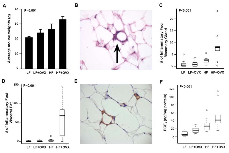
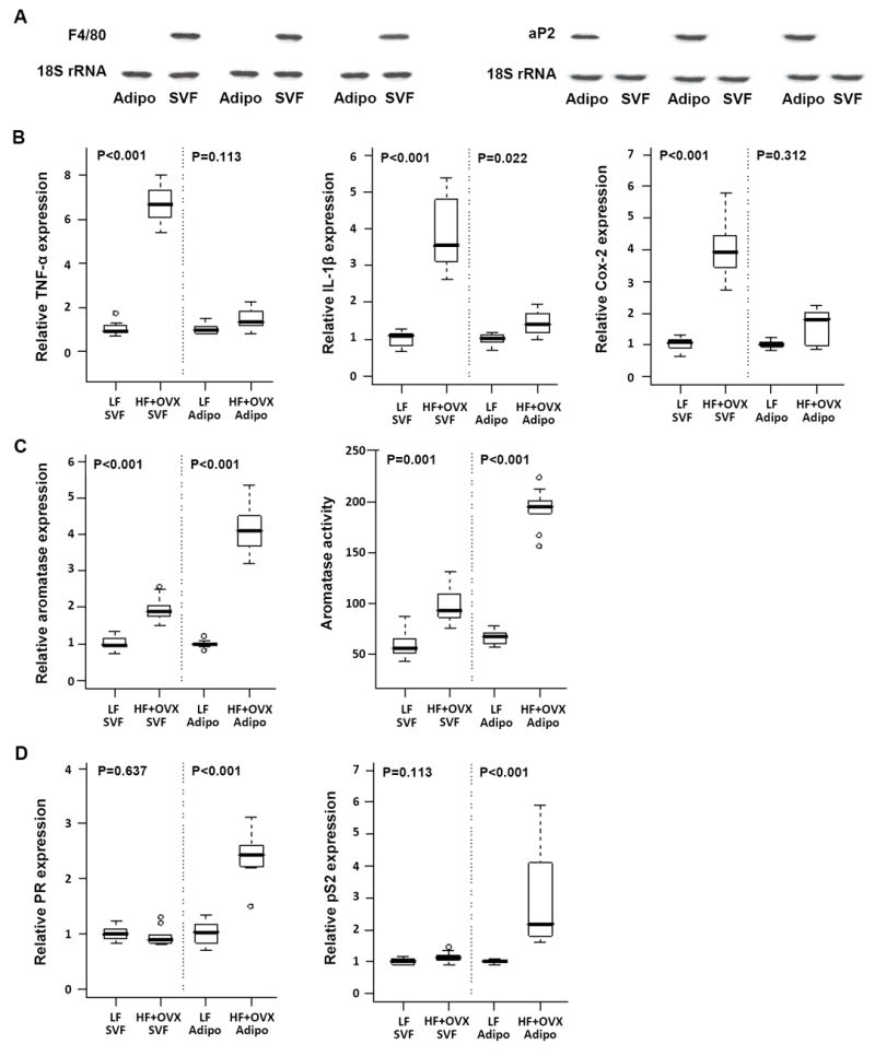
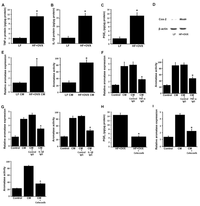
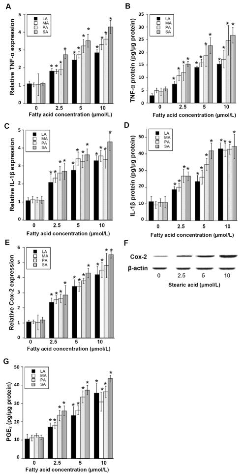
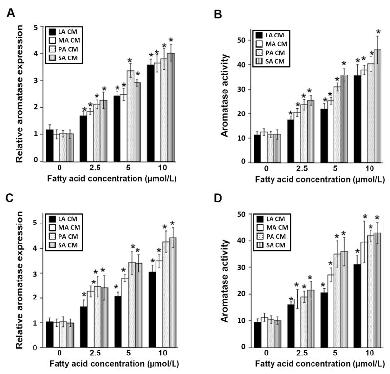
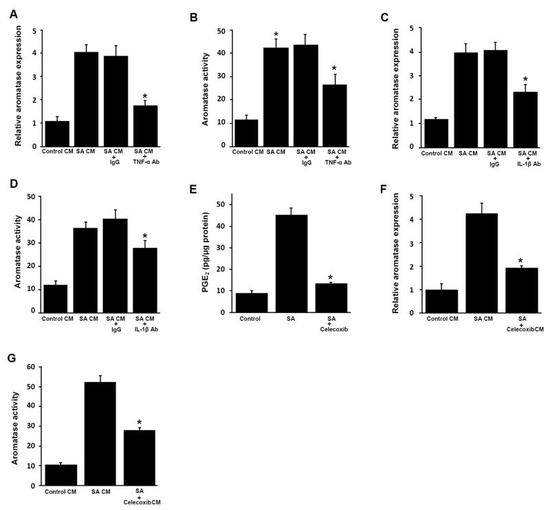
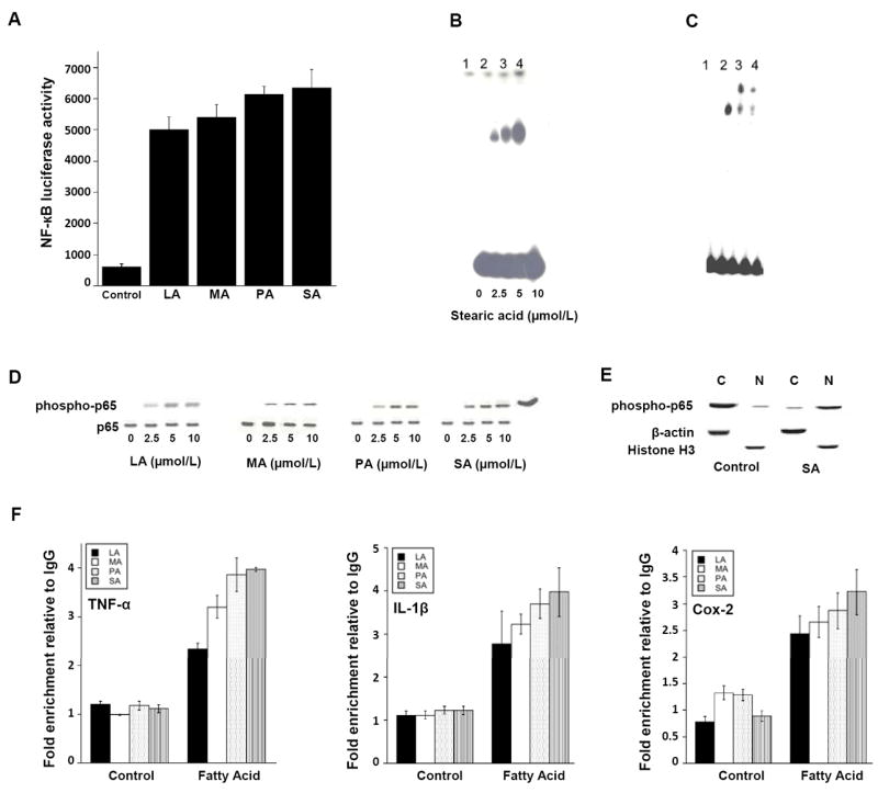
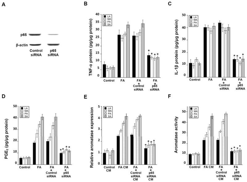
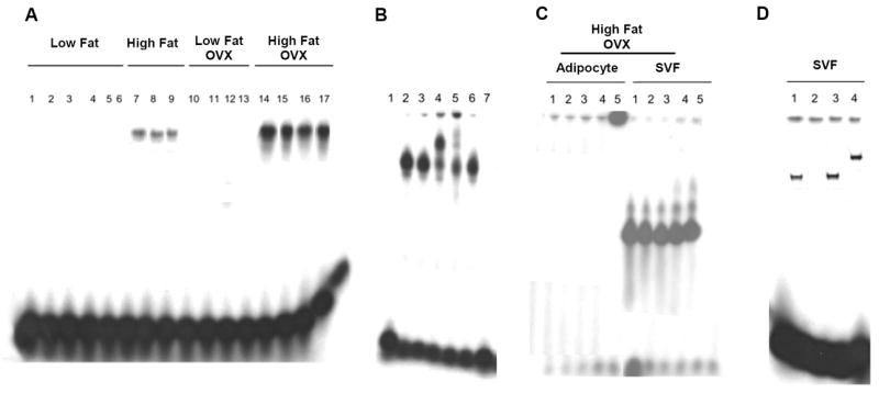
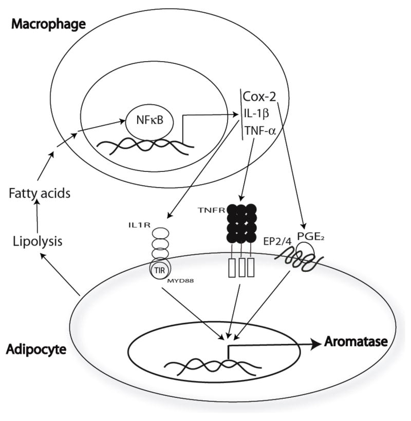
Elevated circulating estrogen levels are associated with increased risk of breast cancer in obese postmenopausal women. Following menopause, the biosynthesis of estrogens through CYP19 (aromatase)-mediated metabolism of androgen precursors occurs primarily in adipose tissue, and the resulting estrogens are then secreted into the systemic circulation. The potential links between obesity, inflammation, and aromatase expression are unknown. In both dietary and genetic models of obesity, we observed necrotic adipocytes surrounded by macrophages forming crown-like structures (CLS) in the mammary glands and visceral fat. The presence of CLS was associated with activation of NF-κB and increased levels of proinflammatory mediators (TNF-α, IL-1β, Cox-2), which were paralleled by elevated levels of aromatase expression and activity in the mammary gland and visceral fat of obese mice. Analyses of the stromal-vascular and adipocyte fractions of the mammary gland suggested that macrophage-derived proinflammatory mediators induced aromatase and estrogen-dependent gene expression (PR, pS2) in adipocytes. Saturated fatty acids, which have been linked to obesity-related inflammation, stimulated NF-κB activity in macrophages leading to increased levels of TNF-α, IL-1β, and Cox-2, each of which contributed to the induction of aromatase in preadipocytes. The discovery of the obesity → inflammation → aromatase axis in the mammary gland and visceral fat and its association with CLS may provide insight into mechanisms underlying the increased risk of hormone receptor-positive breast cancer in obese postmenopausal women, the reduced efficacy of aromatase inhibitors in the treatment of breast cancer in these women, and their generally worse outcomes. The presence of CLS may be a biomarker of increased breast cancer risk or poor prognosis.


Conflict of interest statement

Disclosure of Potential Conflicts of Interest Andrew J. Dannenberg is a member of the Scientific Advisory Board of Tragara Pharmaceuticals Inc., a company that is developing a selective COX-2 inhibitor. The other authors disclosed no potential conflicts of interest.

Figures

Fig. 1
Fig. 1
Diet-induced obesity causes inflammation in the mammary gland and visceral fat. Ovary intact or ovariectomized (OVX) female mice (n=10/group) were fed either a low fat (LF) or high fat (HF) diet for 10 weeks. A, Weight of mice in the diet-induced obesity experiment, shown as mean ± SD. A significant difference in average weights was observed across groups (P<0.001, ANOVA). In particular, OVX mice fed a HF diet had a significantly higher average weight compared to the other groups (P.adj<0.001, Tukey’s test). B, Hematoxylin and eosin stained slides were evaluated for the presence of inflammatory foci containing macrophages that surrounded necrotic adipocytes (arrow, 200x). C and D, Box-plots of the number of inflammatory foci in mammary glands and visceral fat of mice in the different treatment groups. Significant differences were observed across the four experimental groups for both tissue types (P<0.001, Kruskal-Wallis test). In pair-wise comparisons, OVX mice fed the HF diet showed significantly greater number of inflammatory foci in mammary gland compared with those in the LF diet groups (P.adj=0.01, Wilcoxon rank sum test, P values were adjusted for multiple comparisons with Bonferroni method), and in visceral fat compared with those in the other groups (P.adj<=0.01). E, Inflammatory foci contain F4/80-positive macrophages that formed a typical crown around adipocytes (200x). F, Box-plots of PGE2 levels in mammary glands of mice in the different treatment groups. Significant differences were observed across the four experimental groups (P<0.001, Kruskal-Wallis test). In pair-wise comparisons, OVX mice fed the HF diet showed significantly greater PGE2 levels compared with those in the LF diet groups (P.adj=0.01, Wilcoxon rank sum test, P values were adjusted for multiple comparisons with Bonferroni method).
Fig. 2
Fig. 2
Diet-induced obesity is associated with increased levels of pro-inflammatory mediators and aromatase in the mammary gland and visceral fat. Ovary intact or ovariectomized (OVX) female mice (n=10/group) were fed either a low fat (LF) or high fat (HF) diet for 10 weeks. Real-time PCR was carried out on RNA isolated from the mammary gland (n=10/group) and visceral fat (n=10/group) of mice in each of the four groups. Box-plots of TNF-α, IL-1β and Cox-2 mRNA expression in mammary glands (A-C) and visceral fat (F-H) are shown. Significant differences were observed across the four experimental groups for each pro-inflammatory mediator (P<0.005). In pair-wise comparisons, OVX mice fed a HF diet showed significantly higher expression of the three genes in mammary gland and visceral fat compared with mice in the LF or LF + OVX groups (P.adj<=0.02). Box-plots of relative aromatase mRNA levels and activity in mammary glands (D, E) and visceral fat (I, J) of mice in each of the four treatment groups are shown. Significant differences were observed across the four experimental groups for aromatase expression and activity (P<0.001). In pair-wise comparisons, OVX mice fed a HF diet had significantly higher levels of aromatase activity (femtomoles/μg protein/hour) and mRNA expression in mammary gland and visceral fat compared with those in the other groups (P.adj ≤ 0.05).
Fig. 3
Fig. 3
Diet-induced obesity is associated with elevated levels of pro-inflammatory mediators and aromatase in different compartments of the mammary gland. Adipose (Adipo) and stromal-vascular fractions (SVF) were prepared from mammary glands of ovary intact mice fed a low fat (LF) diet or mice that were subjected to ovariectomy (OVX) and fed a high fat (HF) diet for 10 weeks. A, RNA from SVF and adipose fractions was isolated from three mammary glands and subjected to northern blotting. Blots were probed for F4/80 and aP2 as indicated. B-D, Real-time PCR was used to quantify mRNA levels for TNF-α, IL-1β, Cox-2, aromatase, PR and pS2. B, Box-plots of the TNF-α, IL-1β and Cox-2 mRNA levels in the SVF and the adipose fractions of mammary glands (n=10/group) are shown. In comparison to lean mice (LF), obese mice (HF+OVX) showed significantly higher expression levels of all three pro-inflammatory mediators in the SVF but not in the adipose fraction of the mammary gland (P<0.001). C, Box-plots of relative aromatase mRNA levels and aromatase activity in the SVF and adipose fractions of mammary glands (n=10/group) are shown. Aromatase activity is expressed as femtomoles/μg protein/hour. D, Relative progesterone receptor (PR) and pS2 mRNA expression in the SVF and adipose fractions of the mammary gland (n=10/group) are shown.
Fig. 4
Fig. 4
Pro-inflammatory mediators derived from the stromal-vascular fraction of the mammary gland of obese mice induce aromatase in preadipocytes. SVF cells derived from the mammary gland of mice in the LF and HF+OVX groups were grown overnight in DMEM. Conditioned medium (CM) was then collected and analyzed or used to treat 3T3-LI preadipocytes. Levels of TNF-α (A), IL-1β (B) and PGE2 (C) in CM were determined by enzyme immunoassay. D, Cox-2 protein abundance was determined by immunoblotting of whole cell lysates. β-actin was used as a loading control. E, 3T3-L1 cells were treated with CM derived from the mammary gland SVF of LF or HF + OVX mice for 24 hours. Relative aromatase mRNA levels and aromatase activity were then determined. F and G, neutralizing either TNF-α (F) or IL-1β (G) suppressed the ability of CM derived from HF + OVX SVF to induce aromatase in 3T3-L1 cells. In both F and G, the bar labeled Control represents 3T3-L1 cells that received DMEM medium alone; the bar labeled CM represents 3T3-L1 cells that received CM derived from HF + OVX SVF. Additionally, CM from HF + OVX SVF was incubated with control IgG, TNF-α IgG or IL-1β IgG overnight at 4°C to neutralize TNF-α (F) and IL-1β (G), respectively. 3T3-L1 cells were then treated as indicated for 24 hours prior to measurements of relative aromatase mRNA levels and aromatase activity. H, SVF cells derived from the mammary gland of mice in the HF+OVX group were grown overnight. The cells were then treated with fresh DMEM containing vehicle (HF + OVX) or 5 μM celecoxib (HF+OVX+celecoxib) for 6 hours. CM was collected and analyzed for PGE2 levels (H) or used to treat 3T3-LI preadipocytes (I). I, inhibiting Cox-2 in SVF cells derived from HF + OVX mammary gland attenuated the ability of CM to induce aromatase in 3T3-L1 cells. The bar labeled Control represents 3T3-L1 cells that received DMEM medium; the bar labeled CM represents 3T3-L1 cells that received CM derived from HF + OVX SVF; the bar labeled CM + celecoxib represents 3T3-L1 cells that received CM derived from HF + OVX SVF treated with 5 μM celecoxib. 3T3-L1 cells were treated as indicated for 24 hours prior to measurements of relative aromatase mRNA levels and aromatase activity. Aromatase activity is expressed as femtomoles/μg protein/minute. Columns, means (n=3); bars, SD. *P< 0.05.
Fig. 5
Fig. 5
Treatment of macrophages with saturated fatty acids causes dose-dependent induction of pro-inflammatory mediators. THP-1 cells were treated with the indicated concentration of saturated fatty acid (LA, lauric acid; MA, myristic acid; PA, palmitic acid; SA, stearic acid) for 24 hours. Real-time PCR was used to quantify TNF-α, IL-1β and Cox-2 mRNAs (A, C, E). Levels of TNF-α protein, IL-1β protein and PGE2 in the culture medium were determined by enzyme immunoassay (B, D, G). Cox-2 protein abundance was determined by immunoblotting of whole cell lysates. β-actin was used as a loading control (F). Columns, means (n=6); bars, SD. *P < 0.05.
Fig. 6
Fig. 6
Conditioned medium from saturated fatty acid-treated macrophages induces aromatase in preadipocytes. THP-1 cells or human blood monocyte-derived macrophages were treated with 0-10 μmol/L lauric acid (LA), myristic acid (MA), palmitic acid (PA) or stearic acid (SA) as detailed in the Materials and Methods to generate conditioned medium (CM). Conditioned medium was then used to treat preadipocytes for 24 hours. Panels A and B represent preadipocytes treated with CM derived from THP-1 cells. Panels C and D represent preadipocytes treated with CM derived from human blood monocyte-derived macrophages. Aromatase mRNA (A, C) and activity levels (femtomoles/μg protein/minute) (B, D) were determined. Columns, means (n=6); bars, SD. *P < 0.05.
Fig. 7
Fig. 7
Pro-inflammatory mediators in conditioned medium of stearic acid-treated macrophages induce aromatase in preadipocytes. A-D, THP-1 cells were treated with vehicle or 10 μmol/L stearic acid (SA) as detailed in the Materials and Methods to generate conditioned medium (CM). As indicated, CM from SA-treated cells was then incubated with neutralizing antibodies (Ab) to TNF-α, IL-1β or control IgG overnight at 4°C. E-G, THP-1 cells were treated with vehicle, SA or SA and 5 μmol/L celecoxib for 12 hours. Following treatment, the medium was removed and cells were washed. Subsequently, fresh medium was added for 24 hours to generate CM. Panel E indicates that celecoxib suppressed SA-mediated induction of PGE2 production. In A-D, F and G, preadipocytes were treated with THP-1 cell-derived CM for 24 hours prior to measurements of aromatase mRNA and activity. Aromatase mRNA (A, C, F) and activity levels (B, D, G) were determined in preadipocytes. Activity is expressed as femtomoles/μg protein/minute. Columns, means (n=6); bars, SD. *P < 0.05.
Fig. 8
Fig. 8
Saturated fatty acids activate NF-κB in THP-1 cells. A, cells were transiently transfected with NF-κB-luciferase and pSV-βgal constructs. Cells were then treated with 10 μmol/L lauric acid (LA), myristic acid (MA), palmitic acid (PA) or stearic acid (SA) for 24 hours. Activities represent data that have been normalized to β-galactosidase activity. Columns, mean (n=6); bar, S.D. B and C, 10 μg of nuclear protein was incubated with a 32P-labeled oligonucleotide containing NF-κB binding sites. In B, lanes 1-4 represent binding with nuclear protein from cells treated with the indicated concentration of SA for 1 hour. In C, lanes 1-4 represent binding with nuclear protein from cells treated with vehicle (lane 1) or 10 μmol/L SA (lanes 2-4) for 1 hour. Lanes 2-4 represent nuclear protein incubated with normal IgG (lane 2) or antibodies to p65 (2 μL, lane 3; 1 μL, lane 4). In B and C, the protein-DNA complexes that formed were separated on a 4% polyacrylamide gel. D, cells were treated as indicated with 0-10 μmol/L LA, MA, PA or SA for 30 minutes. The abundance of phospho-p65 and p65 protein in cell lysates was determined by immunoblotting. E, cells were treated with vehicle (control) or 10 μmol/L SA for 30 minutes. The abundance of phospho-p65 was determined by immunoblotting in cytosolic (C) and nuclear (N) preparations. β-actin and histone H3 represent cytosolic and nuclear markers, respectively. F, cells were treated with vehicle (Control) or 10 μmol/L LA, MA, PA, SA for 3 hours. ChIP assays were performed. Chromatin fragments were immunoprecipitated with antibodies against phospho-p65 and the TNF-α, IL-1β and Cox-2 promoters were amplified by real-time PCR. DNA sequencing was carried out, and the PCR products were confirmed to be correct promoters. These promoters were not detected when normal IgG was used or when antibody was omitted from the immunoprecipitation step (data not shown). Means ± S.D. are shown; n=3.
Fig. 9
Fig. 9
Silencing of p65 inhibits saturated fatty acid (FA)-mediated induction of pro-inflammatory mediators and aromatase. A, THP-1 cells were transfected with control siRNA or siRNA to p65. The abundance of p65 protein was determined by immunoblotting in cell lysates. β-actin was used as a loading control. B-D, As indicated, THP-1 cells were untreated or transfected with control siRNA or p65 siRNA. Subsequently, cells were treated with 0 or 10 μmol/L lauric acid (LA), myristic acid (MA), palmitic acid (PA) or stearic acid (SA) as detailed in the Materials and Methods to generate conditioned medium (CM). Enzyme immunoassays were used to quantify levels of TNF-α (B), IL-1β (C) and PGE2 (D) in the CM. In E and F, preadipocytes were treated with THP-1 cell-derived CM for 24 hours prior to measurements of aromatase mRNA and activity. Activity is expressed as femtomoles/μg protein/minute. Columns, means (n=6); bars, SD. *P < 0.05.
Fig. 10
Fig. 10
NF-kB is activated in the stromal-vascular fraction of the mammary gland of obese mice. A-D, 10 μg of nuclear protein was incubated with a 32P-labeled oligonucleotide containing NF-κB binding sites. A, binding of nuclear protein from unfractionated mammary glands of 17 mice in the indicated treatment groups. B, lanes 1-7 represent binding of nuclear protein from mammary glands in the High Fat + OVX group. Lane 1, binding reaction without nuclear protein; lane 2, binding of nuclear protein to labeled oligonucleotide; lanes 3-5, nuclear protein incubated with normal IgG (lane 3) or phospho-p65 antibody (2μL, lane 4; 1 μL, lane 5); lanes 6 and 7, nuclear protein was incubated with labeled oligonucleotide and a 10X excess (lane 6) or 50X excess of cold probe (lane 7). C, binding of nuclear protein isolated from the adipocyte or stromal-vascular (SVF) fractions of the mammary gland of 5 mice in the High Fat + OVX group. D, binding of nuclear protein from SVF isolated from a mammary gland in the High Fat + OVX group. Lane 1, binding of nuclear protein to labeled oligonucleotide; lane 2, nuclear protein was incubated with labeled oligonucleotide and a 50X excess of cold probe; lanes 3 and 4, nuclear protein incubated with normal IgG (lane 3) or 2μL of phospho-p65 antibody (lane 4). In A-D, the protein-DNA complexes that formed were separated on a 4% polyacrylamide gel.
Fig. 11
Fig. 11
Paracrine interactions between adipocytes and macrophages can explain the elevated levels of aromatase in the mammary gland and visceral fat of obese mice. In obesity, lipolysis is increased resulting in increased concentrations of free fatty acids. Saturated fatty acids trigger the activation of NF-κB in macrophages resulting in enhanced production of pro-inflammatory mediators (PGE2, TNF-α, IL-1β). Each of these pro-inflammatory mediators contributes to the induction of aromatase in preadipocytes and adipocytes.

Comment in

References

    1. Calle EE, Kaaks R. Overweight, obesity and cancer: epidemiological evidence and proposed mechanisms. Nat Rev Cancer. 2004;4:579–91. - PubMed
    1. Santen RJ, Brodie H, Simpson ER, Siiteri PK, Brodie A. History of aromatase: saga of an important biological mediator and therapeutic target. Endocr Rev. 2009;30:343–75. - PubMed
    1. Lorincz AM, Sukumar S. Molecular links between obesity and breast cancer. Endocr Relat Cancer. 2006;13:279–92. - PubMed
    1. Cleary MP, Grossmann ME. Minireview: Obesity and breast cancer: the estrogen connection. Endocrinology. 2009;150:2537–42. - PMC - PubMed
    1. Key TJ, Appleby PN, Reeves GK, Roddam A, Dorgan JF, Longcope C, et al. Body mass index, serum sex hormones, and breast cancer risk in postmenopausal women. J Natl Cancer Inst. 2003;95:1218–26. - PubMed

Publication types