

δ-tocopherol is more active than α - or γ -tocopherol in inhibiting lung tumorigenesis in vivo
- PMID: 21372040
- PMCID: PMC3071153
- DOI: 10.1158/1940-6207.CAPR-10-0130
δ-tocopherol is more active than α - or γ -tocopherol in inhibiting lung tumorigenesis in vivo
Abstract
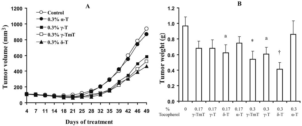
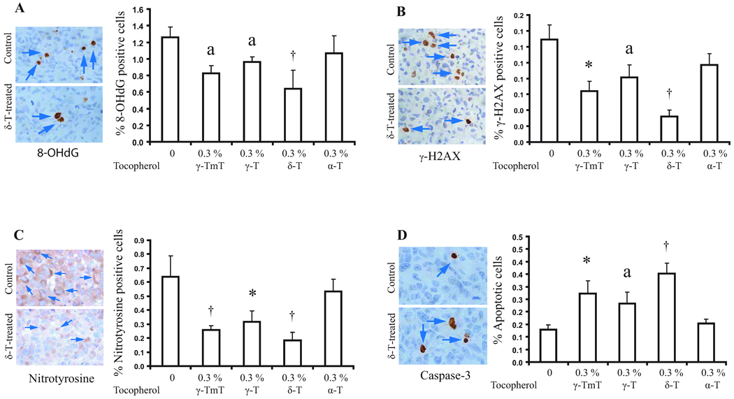
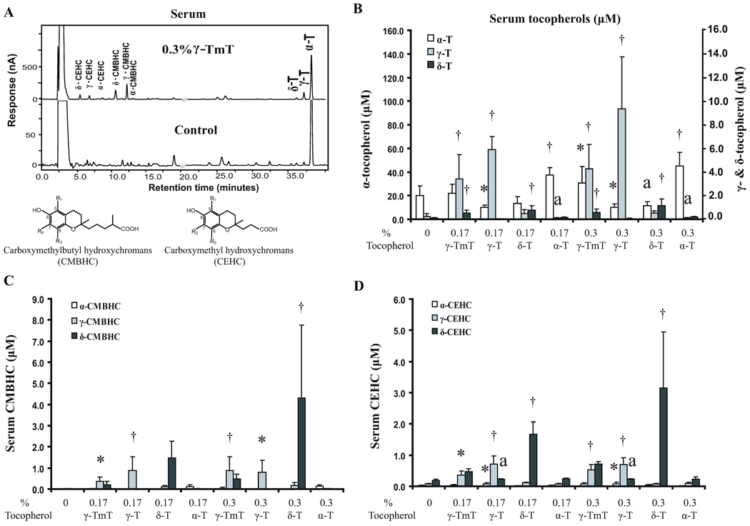
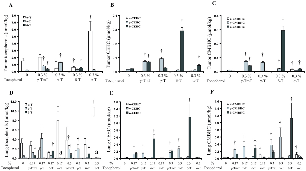
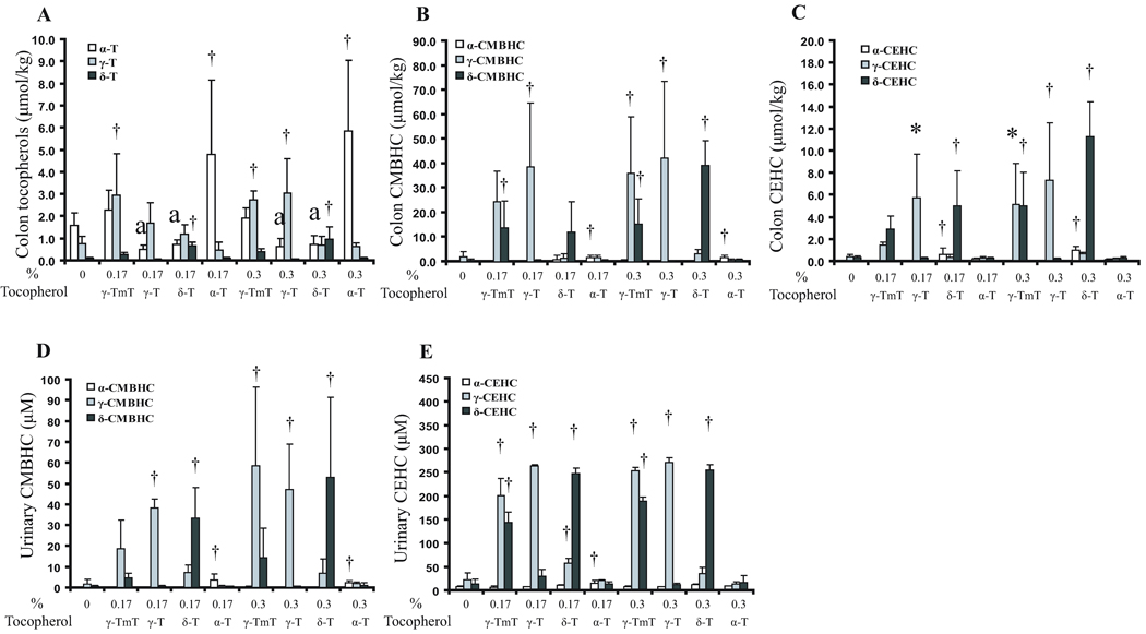
In contrast to strong epidemiologic, preclinical, and secondary clinical evidence for vitamin E (tocopherols) in reducing cancer risk, large-scale clinical cancer-prevention trials of α-tocopherol have been negative. This vexing contrast helped spur substantial preclinical efforts to better understand and improve the antineoplastic activity of tocopherol through, for example, the study of different tocopherol forms. We previously showed that the γ-tocopherol-rich mixture (γ-TmT) effectively inhibited colon and lung carcinogenesis and the growth of transplanted lung-cancer cells in mice. We designed this study to determine the relative activities of different forms of tocopherol in a xenograft model, comparing the anticancer activities of δ-tocopherol with those of α- and γ-tocopherols. We subcutaneously injected human lung cancer H1299 cells into NCr nu/nu mice, which then received α-, γ-, or δ-tocopherol or γ-TmT in the diet (each at 0.17% and 0.3%) for 49 days. δ-Tocopherol inhibited tumor growth most strongly. γ-Tocopherol and γ-TmT (at 0.3%) also inhibited growth significantly, but α-tocopherol did not. δ-Tocopherol also effectively decreased oxidative DNA damage and nitrotyrosine formation and enhanced apoptosis in tumor cells; again, γ-tocopherol also was active in these regards but less so, and α-tocopherol was not. Each supplemented diet increased serum levels of its tocopherol - up to 45 μmol/L for α-tocopherol, 9.7 μmol/L for γ-tocopherol, and 1.2 μmol/L for δ-tocopherol; dietary γ- or δ-tocopherol, however, decreased serum α-tocopherol levels, and dietary α-tocopherol decreased serum levels of γ-tocopherol. Each dietary tocopherol also increased its corresponding side-chain-degradation metabolites, with concentrations of δ-tocopherol metabolites greater than γ-tocopherol and far greater than α-tocopherol metabolites in serum and tumors. This study is the first in vivo assessment of δ-tocopherol in tumorigenesis and shows that δ-tocopherol is more active than α- or γ-tocopherol in inhibiting tumor growth, possibly through trapping reactive oxygen and nitrogen species and inducing apoptosis; δ-tocopherol metabolites could contribute significantly to these results.
Conflict of interest statement
Disclosure of Potential Conflicts of Interest
No Potential conflicts of interest were disclosed.
Figures
References
-
- Traber MG. Vitamin E. In: Bowman BA, M RR, editors. Present Knowledge in Nutrition. Washington DC: ILSI Press; 2006. pp. 211–219.
-
- Taylor PR, Qiao YL, Abnet CC, et al. Prospective study of serum vitamin E levels and esophageal and gastric cancers. J Natl Cancer Inst. 2003;95:1414–1416. - PubMed
-
- Greenwald P, Clifford CK, Milner JA. Diet and cancer prevention. Eur J Cancer. 2001;37:948–965. - PubMed
-
- Knekt P, Aromaa A, Maatela J, et al. Vitamin E and cancer prevention. Am J Clin Nutr. 1991;53:283S–286S. - PubMed
Publication types
MeSH terms
Substances
Grants and funding
LinkOut - more resources
Full Text Sources
Other Literature Sources
Medical
Research Materials
