. 2011 May 13;286(19):17005-14.
doi: 10.1074/jbc.M110.216267. Epub 2011 Mar 3.

Reduced utilization of selenium by naked mole rats due to a specific defect in GPx1 expression

Affiliations

Reduced utilization of selenium by naked mole rats due to a specific defect in GPx1 expression

Marina V Kasaikina et al. J Biol Chem. .

Abstract

Naked mole rat (MR) Heterocephalus glaber is a rodent model of delayed aging because of its unusually long life span (>28 years). It is also not known to develop cancer. In the current work, tissue imaging by x-ray fluorescence microscopy and direct analyses of trace elements revealed low levels of selenium in the MR liver and kidney, whereas MR and mouse brains had similar selenium levels. This effect was not explained by uniform selenium deficiency because methionine sulfoxide reductase activities were similar in mice and MR. However, glutathione peroxidase activity was an order of magnitude lower in MR liver and kidney than in mouse tissues. In addition, metabolic labeling of MR cells with (75)Se revealed a loss of the abundant glutathione peroxidase 1 (GPx1) band, whereas other selenoproteins were preserved. To characterize the MR selenoproteome, we sequenced its liver transcriptome. Gene reconstruction revealed standard selenoprotein sequences except for GPx1, which had an early stop codon, and SelP, which had low selenocysteine content. When expressed in HEK 293 cells, MR GPx1 was present in low levels, and its expression could be rescued neither by removing the early stop codon nor by replacing its SECIS element. In addition, GPx1 mRNA was present in lower levels in MR liver than in mouse liver. To determine if GPx1 deficiency could account for the reduced selenium content, we analyzed GPx1 knock-out mice and found reduced selenium levels in their livers and kidneys. Thus, MR is characterized by the reduced utilization of selenium due to a specific defect in GPx1 expression.


Figures

FIGURE 1.
FIGURE 1.
XFM and ICP-MS analysis of selenium in mouse and MR tissues. Shown are XFM scans of mouse and MR livers (A) and testes (B). Each element and its maximum and minimum threshold values are given above each image in ng/cm2. The rainbow-colored scale bar relates to the signal intensity measured as ng/cm2 in each spot, with dark pixels representing areas of low concentration and a gradient to bright pixels depicting increasing concentrations. A scale bar is shown below the elemental maps. C, selenium was analyzed by ICP-MS in mouse and MR tissues. Values are means ± S.D. (error bars). Organs in which trace elements were analyzed are shown below each panel.
FIGURE 2.
FIGURE 2.
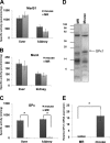
MR tissue extracts have low GPx1 expression and activity. Total MsrB (A), MsrA (B), and GPx (C) activities were measured in the indicated mouse and MR tissues. D, primary splenocytes derived from MR and mice were metabolically labeled with 75Se, and protein extracts were analyzed by SDS-PAGE followed by autoradiography. The band corresponding to GPx1 is shown with an arrow on the right. E, relative expression of GPx1 mRNA in MR and mouse liver (*, p < 0.05). Error bars, S.D.
FIGURE 3.
FIGURE 3.
Transcriptome analysis and characterization of the MR selenoproteome. A, length distribution of assembled contigs of the MR liver transcriptome. B, selenoproteins identified in MR. The human selenoproteome is used as a reference to represent the MR selenoproteome. Selenoproteins identified by sequencing the MR liver transcriptome are highlighted. The location of Sec residues is indicated by a red line. Seven Sec residues were found in SelP. GPx1 has an early termination codon (see “Results”). C, schematic representation of MR GPx1 in comparison with mouse GPx1.
FIGURE 4.
FIGURE 4.
GPx1 is poorly expressed in mammalian cells. A, schematic representation of MR and mouse GPx1 mutant constructs. Positions of Sec and glutamine (CAG) codons, the stop signal (TAG), and the SECIS element in the 3′-UTR are shown. B, mRNA levels were assessed by real-time PCR and normalized to GFP expressed from the same vector. Results are given ± S.D. (error bars). C, MR and mouse Myc-GPx1 constructs were transfected into HEK 293 cells. Cells were labeled with 75Se, followed by SDS-PAGE and autoradiography (top). Migration of ectopically expressed GPx1 is shown on the left. The same lysates were probed with anti-Myc and anti-β-actin antibodies (two bottom panels). Myc-GPx1 mutants were also immunoprecipitated with anti-Myc antibodies, followed by PhosphorImager analysis of the 75Se-labeled GPx1. D, mouse and MR constructs were transfected into HEK 293 cells followed by treatment of cells with 10 μm MG132 for 12 h. Cells were labeled with 75Se, followed by protein analysis by SDS-PAGE and autoradiography (top). The same membrane was stained with Myc and GFP antibodies. E, expression of mouse and MR cysteine mutants of GPx1 in HEK 293 cells was analyzed by Western blotting. IP, immunoprecipitation; WB, Western blotting.
FIGURE 5.
FIGURE 5.
SECIS element does not decrease GPx1 expression level. A, alignment of mammalian GPx1 SECIS elements. Nucleotides critical for the SECIS function are underlined. B, MR GPx1 SECIS element as predicted by SECISearch. Functional sites are shown in boldface type (24). C, mouse GPx1 SECIS element. Functional sites are shown in boldface type. D, mouse and MR GPx1 coding sequences were cloned into pSelExpress1 containing an efficient eukaryotic SECIS element and expressed in HEK 293 cells. Cells were labeled with 75Se, followed by SDS-PAGE, autoradiography, and Western blotting with Myc antibodies. Coomassie staining is a loading control. E, coding sequences of mouse GPx1 containing the MR GPx1 3′-UTR (M-long-MR SECIS) and coding sequence of MR GPx1 containing mouse GPx1 3′-UTR (MR-short-M SECIS) were expressed in HEK 293 cells. Cells were labeled with 75Se, followed by SDS-PAGE, autoradiography, and Western blotting with Myc and GFP antibodies. WB, Western blot.
FIGURE 6.
FIGURE 6.
Reduction in selenium levels in tissues of GPx1 KO mice. Selenium was determined by ICP-MS in tissues from wild type (WT) and GPx1 knock-out (KO) mice. Values are means ± S.D. (error bars). Organs in which selenium was analyzed are shown below each panel (**, p < 0.005; *, p < 0.05).

References

    1. Buffenstein R. (2005) J. Gerontol. A Biol. Sci. Med. Sci. 60, 1369–1377 - PubMed
    1. Buffenstein R. (2008) J. Comp. Physiol. B 178, 439–445 - PubMed
    1. Edrey Y. H., Park T. J., Kang H., Biney A., Buffenstein R. (2011) Exp. Gerontol. 46, 116–123 - PubMed
    1. McMullen C. A., Andrade F. H., Crish S. D. (2010) Anat. Rec. 293, 918–923 - PMC - PubMed
    1. Park T. J., Lu Y., Jüttner R., Smith E. S., Hu J., Brand A., Wetzel C., Milenkovic N., Erdmann B., Heppenstall P. A., Laurito C. E., Wilson S. P., Lewin G. R. (2008) PLoS Biol. 6, e13. - PMC - PubMed

Publication types

LinkOut - more resources