

Echocardiographic speckle-tracking based strain imaging for rapid cardiovascular phenotyping in mice
- PMID: 21372284
- PMCID: PMC3376717
- DOI: 10.1161/CIRCRESAHA.110.239574
Echocardiographic speckle-tracking based strain imaging for rapid cardiovascular phenotyping in mice
Abstract
Rationale: High-sensitivity in vivo phenotyping of cardiac function is essential for evaluating genes of interest and novel therapies in small animal models of cardiovascular disease. Transthoracic echocardiography is the principal method currently used for assessing cardiac structure and function; however, standard echocardiographic techniques are relatively insensitive to early or subtle changes in cardiac performance, particularly in mice.
Objective: To develop and validate an echocardiographic strain imaging methodology for sensitive and rapid cardiac phenotyping in small animal models.
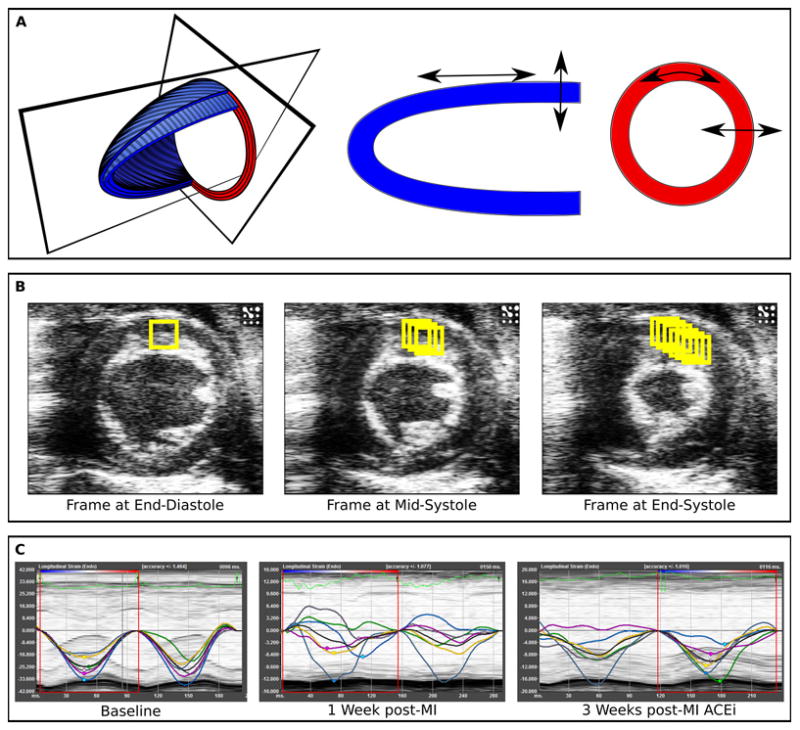
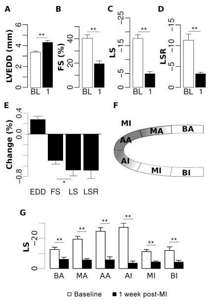
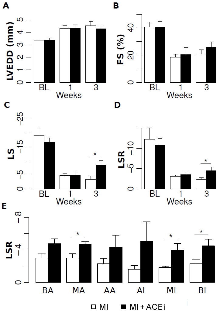
Methods and results: Herein, we describe a modified echocardiographic technique that uses speckle-tracking based strain analysis for the noninvasive evaluation of cardiac performance in adult mice. This method is found to be rapid, reproducible, and highly sensitive in assessing both regional and global left ventricular (LV) function. Compared with conventional echocardiographic measures of LV structure and function, peak longitudinal strain and strain rate were able to detect changes in adult mouse hearts at an earlier time point following myocardial infarction and predicted the later development of adverse LV remodeling. Moreover, speckle-tracking based strain analysis was able to clearly identify subtle improvement in LV function that occurred early in response to standard post-myocardial infarction cardiac therapy.
Conclusions: Our results highlight the utility of speckle-tracking based strain imaging for detecting discrete functional alterations in mouse models of cardiovascular disease in an efficient and comprehensive manner. Echocardiography speckle-tracking based strain analysis represents a method for relatively high-throughput and sensitive cardiac phenotyping, particularly in evaluating emerging cardiac agents and therapies in mice.
Figures
References
-
- Christensen G, Wang Y, Chien KR. Physiological assessment of complex cardiac phenotypes in genetically engineered mice. Am J Physiol. 1997;272:H2513–2524. - PubMed
-
- Hanton G. Preclinical cardiac safety assessment of drugs. Drugs R D. 2007;8:213–228. - PubMed
-
- Marwick TH, Raman SV, Carrio I, Bax JJ. Recent developments in heart failure imaging. JACC Cardiovasc Imaging. 2010;3:429–439. - PubMed
-
- Stanton T, Marwick TH. Assessment of subendocardial structure and function. JACC Cardiovasc Imaging. 2010;3:867–875. - PubMed
-
- Lang RM, Bierig M, Devereux RB, Flachskampf FA, Foster E, Pellikka PA, Picard MH, Roman MJ, Seward J, Shanewise JS, Solomon SD, Spencer KT, Sutton MS, Stewart WJ. Recommendations for chamber quantification: a report from the American Society of Echocardiography’s Guidelines and Standards Committee and the Chamber Quantification Writing Group, developed in conjunction with the European Association of Echocardiography, a branch of the European Society of Cardiology. J Am Soc Echocardiogr. 2005;18:1440–1463. - PubMed
Publication types
MeSH terms
Grants and funding
LinkOut - more resources
Full Text Sources
Other Literature Sources
Medical
