Review
. 2011 Mar;75(1):133-91.
doi: 10.1128/MMBR.00015-10.

Oxidative stress resistance in Deinococcus radiodurans

Affiliations
Review

Oxidative stress resistance in Deinococcus radiodurans

Dea Slade et al. Microbiol Mol Biol Rev. 2011 Mar.

Abstract

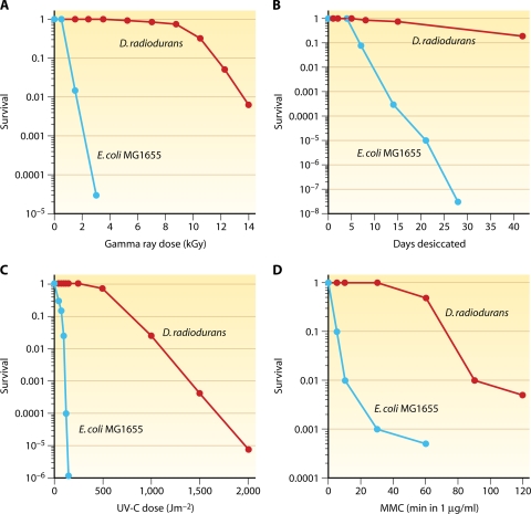
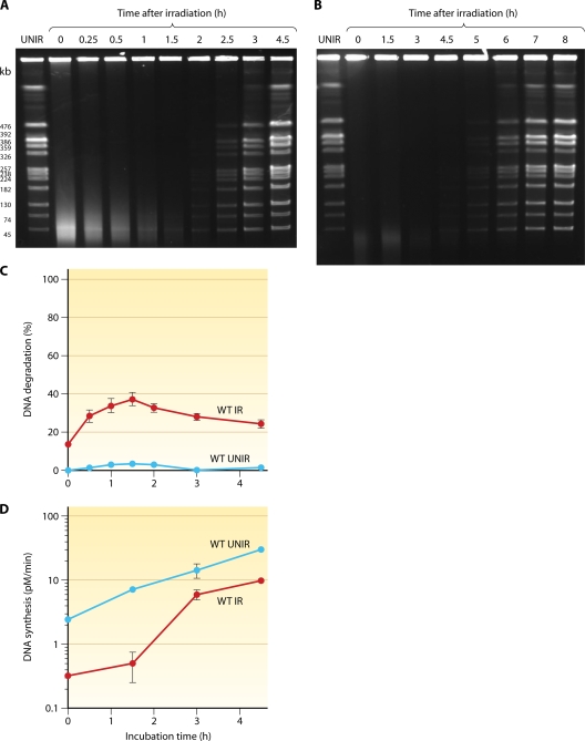
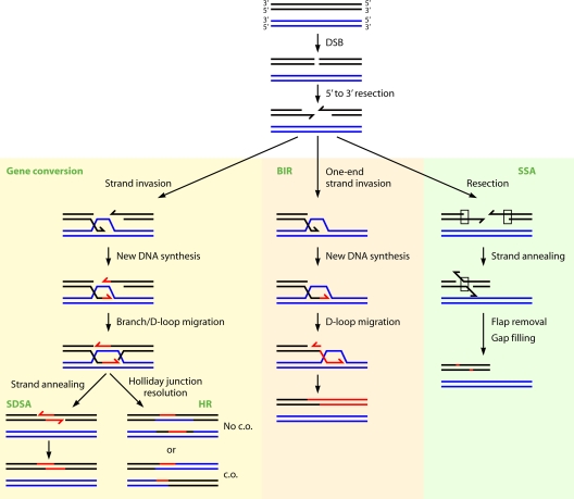
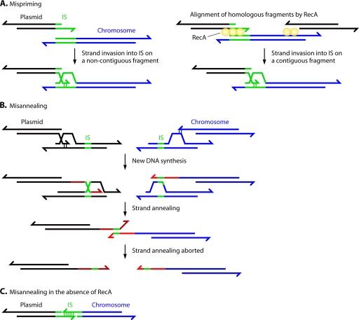
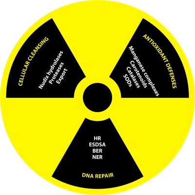
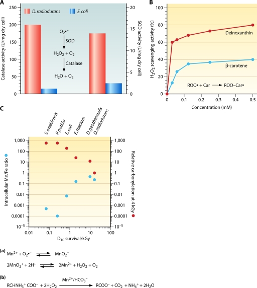
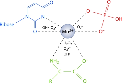
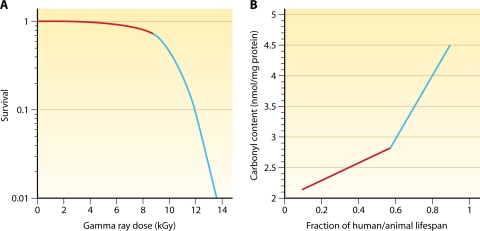
Deinococcus radiodurans is a robust bacterium best known for its capacity to repair massive DNA damage efficiently and accurately. It is extremely resistant to many DNA-damaging agents, including ionizing radiation and UV radiation (100 to 295 nm), desiccation, and mitomycin C, which induce oxidative damage not only to DNA but also to all cellular macromolecules via the production of reactive oxygen species. The extreme resilience of D. radiodurans to oxidative stress is imparted synergistically by an efficient protection of proteins against oxidative stress and an efficient DNA repair mechanism, enhanced by functional redundancies in both systems. D. radiodurans assets for the prevention of and recovery from oxidative stress are extensively reviewed here. Radiation- and desiccation-resistant bacteria such as D. radiodurans have substantially lower protein oxidation levels than do sensitive bacteria but have similar yields of DNA double-strand breaks. These findings challenge the concept of DNA as the primary target of radiation toxicity while advancing protein damage, and the protection of proteins against oxidative damage, as a new paradigm of radiation toxicity and survival. The protection of DNA repair and other proteins against oxidative damage is imparted by enzymatic and nonenzymatic antioxidant defense systems dominated by divalent manganese complexes. Given that oxidative stress caused by the accumulation of reactive oxygen species is associated with aging and cancer, a comprehensive outlook on D. radiodurans strategies of combating oxidative stress may open new avenues for antiaging and anticancer treatments. The study of the antioxidation protection in D. radiodurans is therefore of considerable potential interest for medicine and public health.


Figures

FIG. 1.
FIG. 1.
D. radiodurans cell division on TGY agar at 30°C. (A) Cell division monitored by time-lapse fluorescent microscopy starting from a single diad (see Video S1 in the supplemental material). Images were obtained with a fluorescent microscope (Zeiss Axiovert 200 M). Membranes were stained with FM 4-64 (red), and nucleoids were stained with 4′,6-diamidino-2-phenylindole (DAPI) (blue). Septa are formed perpendicular to the cross wall, separating the two cells in a diad (step 1). Septation creates two new cross walls, and two diads are formed as a result. Perpendicular to the previously formed cross walls, new septa invaginate into four cells with nuclear material in the process of separating into daughter cells (step 2). This stage of cell division represents two tetrads. Complete septation results in four new cross walls that give rise to four diads (step 3). New septa invaginate perpendicular to the previously formed cross walls, giving rise to four tetrads (step 4). The following round of cell division produces eight tetrads (step 5). (B) Phase contrast-image of a microcolony obtained from a single tetrad.
FIG. 2.
FIG. 2.
Sources of energy and building blocks in oxidatively stressed D. radiodurans. (A) Extracellular proteolysis. D. radiodurans contains many secreted proteases and ABC transporters, which provide exogenous amino acids as protein building blocks and peptides as components of manganese complexes. (B) The pentose phosphate pathway (PPP) for the conversion of glucose into dNTPs. Glucose is essential for recovery from oxidative stress; glucose-6-phosphate dehydrogenase (the marker enzyme of PPP) converts glucose into the precursors of dNTPs as DNA building blocks and, possibly, components of Mn complexes. (C) Carbohydrate and polyphosphate granules. Shown is a schematic representation of a D. radiodurans tetrad viewed by electron microscopy (based on data from reference and unpublished data from J. R. Battista). The small light granules contain carbohydrates, while the large dark circles represent polyphosphate granules. Both can be used as sources of energy, while polyphosphates also serve as a phosphate source for the synthesis of nucleic acids and, presumably, for manganese-phosphate complexes.
FIG. 3.
FIG. 3.
Extreme resistance of D. radiodurans to gamma rays (A), desiccation (B), UV-C radiation (100 to 295 nm) (C), and mitomycin C (D) in comparison with E. coli. (Panel B is based on data from references and , panel C is based in part on E. coli data from reference , and panel D is based in part on D. radiodurans data from reference .)
FIG. 4.
FIG. 4.
Kinetics of DNA fragment joining in D. radiodurans after 7 kGy (A) and 14 kGy (B) of gamma rays monitored by PFGE. Samples of unirradiated and irradiated wild-type cells were taken to prepare DNA plugs, which were digested with NotI, generating 12 visible fragments. Lane “UNIR” shows the NotI restriction pattern of DNA from unirradiated cells, lane “0” shows the NotI restriction pattern of DNA from irradiated cells immediately after irradiation, and subsequent lanes show the NotI restriction patterns of DNA from cells at different time points after irradiation, expressed in hours. (C) DNA degradation measured in 3H-prelabeled unirradiated (blue) and 7-kGy-irradiated (red) wild-type (WT) cells. (D) Rate of DNA synthesis in unirradiated (blue) and 7-kGy-irradiated (red) wild-type cells. The rate of DNA synthesis is expressed as the amount of [3H]thymidine (pM) incorporated into DNA per minute. (Panels C and D are modified from reference with permission from Elsevier.)
FIG. 5.
FIG. 5.
Linear relationship between the dose of ionizing radiation and, from top to bottom, the extent of DNA degradation and the delay in DNA synthesis. The delay in DNA synthesis is linearly correlated with a dose of up to 4 kGy, above which it reaches a plateau. (Modified from reference with permission of the publisher and based in part on data from references and .)
FIG. 6.
FIG. 6.
Two-step mechanism of DNA repair in D. radiodurans shattered by ionizing radiation. Several genomic copies of D. radiodurans undergo random DNA double-strand breakage, producing numerous fragments (step 1). The fragmented DNA is recessed in a 5′-to-3′ direction, presumably by RecJ, liberating single-stranded 3′ overhangs (step 2), which, through RecA- and RadA-mediated strand invasion, prime synthesis on overlapping fragments through a migrating D loop (step 3). DNA synthesis is initiated by Pol III (step 4) and elongated by Pol III, with Pol I filling up gaps arising from the excision repair of damaged bases (A), or by Pol I alone (B).Two noncontiguous fragments are linked by convergent elongations on a third “bridging” fragment (step 5). Newly synthesized single strands dissociate from the template (step 6) and anneal to complementary single-stranded extensions, forming dsDNA intermediates (step 7). The flaps are removed (by SbcCD?), and the gaps are filled (by Pol I?) (step 8). Long linear intermediates are joined into circular chromosomes by RecA-dependent crossovers (step 9). (Modified from reference with permission from Elsevier and based in part on data from reference .)
FIG. 7.
FIG. 7.
Model for the processing of DSB ends and the initiation of recombination in D. radiodurans by the RecFOR pathway. UvrD unwinds the ends of DSBs in the 3′-to-5′ direction, and RecJ digests the 5′ end (step 1). 3′ single-stranded tails are coated by SSB proteins (step 2). RecF binds to the ssDNA-dsDNA junction (step 3) and promotes the assembly of the RecOR complex (RecO-to-RecR ratio, 4:2) onto the junctions (step 4). RecOR displaces SSB proteins and loads RecA onto 3′ ssDNA (step 5). (Based on data from references and .)
FIG. 8.
FIG. 8.
Effect of mutations in DNA repair-related genes on the resistance of D. radiodurans to ionizing radiation (A) (based on data from references , , , , , , , , , , , , , , , and 665), UV-C radiation resistance (B) (based on data from references , , , , , , , , , , , and and unpublished data from D. Slade), and MMC resistance (C) (based on data from references , , , , , , , , , , , , and and unpublished data from D. Slade). Genes are classified into three groups according to the sensitivity of their mutants: extremely sensitive genes are depicted in black, highly sensitive genes are depicted in red, and moderately/slightly sensitive genes are depicted in blue. Designations for radiation sensitivity are relative to wild-type D. radiodurans, as even the most sensitive repair-deficient D. radiodurans mutants are more resistant than many bacteria which encode a full complement of DNA repair genes. The compiled data were not obtained under the same experimental conditions.
FIG. 9.
FIG. 9.
Mechanisms of DSB repair involving homologous sequences. DSB repair commences with the exonucleolytic resection of the ends of DSBs in the 5′-to-3′ direction to produce 3′ single-stranded tails. In gene conversion and break-induced replication (BIR), 3′ tails invade a homologous template. Strand invasion creates a D loop, where 3′ ends prime new DNA synthesis. Gene conversion further branches into two different mechanisms. In synthesis-dependent strand annealing (SDSA), newly synthesized DNA strands formed by D-loop migration are displaced from the template and anneal to each other to restore a contiguous chromosome in a noncrossover configuration. In the homologous recombination (HR) model, two Holliday junctions are formed as the D loop created by strand invasion pairs with the other side of the DSB (second-end capture) and the 3′ end of the noninvading strand is extended by DNA synthesis. Two Holliday junctions can branch migrate to enlarge the heteroduplex region. Holliday junctions can be cleaved by a resolvase by cutting either the two noncrossed strands or the two crossed strands. If both Holliday junctions are cleaved in the same way, gene conversion is not associated with crossover (No c.o.), whereas differential cleavage results in crossover (c.o.). BIR occurs when only one end of a DSB is available for recombination. Such one-ended strand invasion results in extensive DNA synthesis, which may extend hundreds of kilobases. Intrachromosomal single-strand annealing (SSA) enables the repair of a DSB that occurs between two flanking homologous regions (direct repeats). Resection produces single-stranded tails in which complementary strands of the duplicated sequence are exposed and can reanneal, resulting in a deletion of the intervening sequence. Interchromosomal SSA occurs between two chromosomal copies or between homologous sequences on different genomic elements (Fig. 10C) and may not result in a deletion. (Based on data from reference .)
FIG. 10.
FIG. 10.
Fidelity of DSB repair in D. radiodurans. (A and B) The fidelity of DSB repair can be compromised by insertion sequences (ISs) at the level of the priming of new DNA synthesis (A) and the annealing of newly synthesized single strands (B). (A) Mispriming could occur between identical ISs present on noncontiguous DNA fragments. RecA may ensure the fidelity of priming by aligning homologous DNA fragments. (B) Misannealing could occur when newly synthesized single strands from noncontiguous fragments contain identical ISs. The fidelity of annealing is ensured via the synthesis of long single-stranded overhangs, which are much longer than the longest IS. (C) Rearrangements observed in the absence of a functional RecA protein may result from the single-strand annealing (SSA) of noncontiguous fragments that share identical ISs.
FIG. 11.
FIG. 11.
Factors contributing to ionizing radiation resistance in D. radiodurans: cellular cleansing, antioxidant defenses, and DNA repair. Cellular cleansing involves the degradation of oxidized nucleotides by Nudix hydrolases, the export of damaged oligonucleotides, and the proteolytic degradation of damaged proteins. The antioxidant defense system consists of nonenzymatic scavengers, such as manganese complexes and carotenoids, and enzymatic scavengers, such as catalases and superoxide dismutases. Within the DNA repair machinery, base excision repair (BER) acts on damaged DNA bases, and nucleotide excision repair (NER) removes damaged nucleotides, while extended synthesis-dependent strand annealing (ESDSA) and homologous recombination (HR) mend DNA double-strand breaks.
FIG. 12.
FIG. 12.
Schematic representation of a protein network (I) and the consequences of increasing carbonylation of proteins (II and III) on its activity. Red dots represent single carbonyl groups.
FIG. 13.
FIG. 13.
Enzymatic (A) and nonenzymatic (B and C) antioxidant defenses in D. radiodurans. (A) D. radiodurans has higher levels of catalase and superoxide dismutase (SOD) activities than does E. coli. SOD converts superoxide ions (O2·) into hydrogen peroxide (H2O2), which is converted by catalase into water and oxygen. (Based on data from reference .) (B) The D. radiodurans major carotenoid, deinoxanthin, has a higher level of H2O2-scavenging activity than does β-carotene. Carotenoids scavenge peroxyl radicals by the radical adduct formation mechanism. (Modified from reference with permission from Elsevier.) (C) A high intracellular Mn/Fe ratio is correlated with a high radiation resistance level and a low protein oxidation (carbonylation) level among bacteria. Divalent manganese ions (Mn2+) can scavenge O2· in complex with phosphates and H2O2 in complex with amino acids and bicarbonate. (Modified from reference with permission of the publisher and based on data from references , , , , and .)
FIG. 14.
FIG. 14.
Manganese-based chemical antioxidants in D. radiodurans. Divalent manganese (Mn2+) complexes scavenge long-lived (O2· and H2O2) and short-lived (OH·) ROS, thereby preventing their interconversion and proliferation in cells. Mn2+ catalytically scavenges superoxide radicals (O2·) in complex with orthophosphate (red). Free amino acids or peptides in complex with Mn2+ and orthophosphate (or bicarbonate) catalytically decompose hydrogen peroxide (H2O2) and scavenge O2· (green). Nucleosides (and their analogs) containing two carbonyl groups separated by an amino group (e.g., uridine) complex with Mn2+-orthophosphate and scavenge O2· (blue). Nucleosides, free amino acids, peptides, and other small organic metabolites stoichiometrically scavenge hydroxyl radicals (OH·). Note that a single Mn2+ cannot bind all three ligands at the same time. It has been proposed that the DNA repair proteins of D. radiodurans work so efficiently because they are protected from ROS by Mn2+ complexes. (Based on data from reference .)
FIG. 15.
FIG. 15.
The shoulder of the survival curve (red) of D. radiodurans to various DNA-damaging agents such as gamma rays (A) is phenomenologically equivalent to the shoulder of the protein oxidation curve (red) in the reproductive stage of the life cycle in humans and animals (B), in that both reflect little oxidative damage. The accumulation of oxidative damage, at high radiation doses in D. radiodurans or during the last one-third of the life span of humans, causes the shoulder of the survival curve to fall and carbonylation levels to rise exponentially. (Modified from reference with permission from Elsevier.)

Similar articles

Cited by

References

    1. Abed, R. M., B. Zein, A. Al-Thukair, and D. de Beer. 2007. Phylogenetic diversity and activity of aerobic heterotrophic bacteria from a hypersaline oil-polluted microbial mat. Syst. Appl. Microbiol. 30:319-330. - PubMed
    1. Reference deleted.
    1. Abreu, I. A., et al. 2008. The kinetic mechanism of manganese-containing superoxide dismutase from Deinococcus radiodurans: a specialized enzyme for the elimination of high superoxide concentrations. Biochemistry 47:2350-2356. - PubMed
    1. Agostini, H. J., J. D. Carroll, and K. W. Minton. 1996. Identification and characterization of uvrA, a DNA repair gene of Deinococcus radiodurans. J. Bacteriol. 178:6759-6765. - PMC - PubMed
    1. Aguilaniu, H., L. Gustafsson, M. Rigoulet, and T. Nystrom. 2001. Protein oxidation in G0 cells of Saccharomyces cerevisiae depends on the state rather than rate of respiration and is enhanced in pos9 but not yap1 mutants. J. Biol. Chem. 276:35396-35404. - PubMed

LinkOut - more resources