. 2011 Apr;12(4):327-33.
doi: 10.1038/embor.2011.19. Epub 2011 Mar 4.

The Drosophila peptidoglycan-recognition protein LF interacts with peptidoglycan-recognition protein LC to downregulate the Imd pathway

Affiliations

The Drosophila peptidoglycan-recognition protein LF interacts with peptidoglycan-recognition protein LC to downregulate the Imd pathway

Nada Basbous et al. EMBO Rep. 2011 Apr.

Abstract

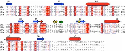
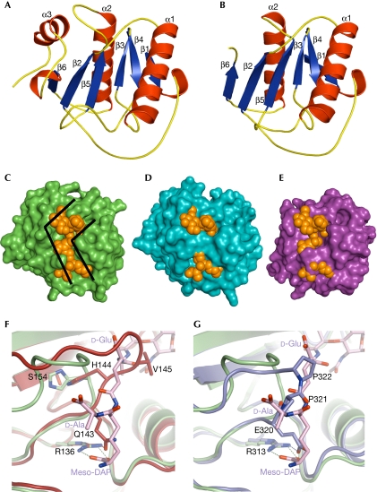
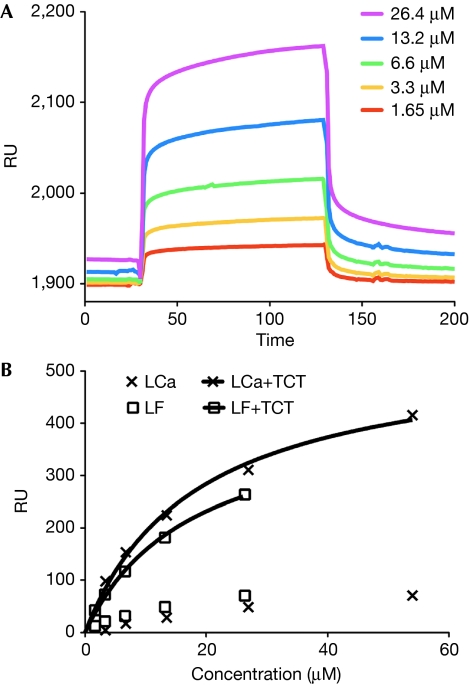
The peptidoglycan (PGN)-recognition protein LF (PGRP-LF) is a specific negative regulator of the immune deficiency (Imd) pathway in Drosophila. We determine the crystal structure of the two PGRP domains constituting the ectodomain of PGRP-LF at 1.72 and 1.94 Å resolution. The structures show that the LFz and LFw domains do not have a PGN-docking groove that is found in other PGRP domains, and they cannot directly interact with PGN, as confirmed by biochemical-binding assays. By using surface plasmon resonance analysis, we show that the PGRP-LF ectodomain interacts with the PGRP-LCx ectodomain in the absence and presence of tracheal cytotoxin. Our results suggest a mechanism for downregulation of the Imd pathway on the basis of the competition between PRGP-LCa and PGRP-LF to bind to PGRP-LCx.


Conflict of interest statement

The authors declare that they have no conflict of interest.

Figures

Figure 1
Figure 1
Sequence alignment of the LFz, LFw, LCx and LCa peptidoglycan-recognition protein domains. Secondary structure elements are indicated above the sequences as blue arrows and red cylinders. Conserved residues are boxed and strictly conserved residues are shown in white with a red background. The conserved arginine residue determinant for the DAP-type specificity is marked with a red star. Important residues that are responsible for the lack of the binding of LFz and LFw to PGN are marked with yellow and green stars, respectively. DAP, diaminopimelic acid; PGN, peptidoglycan; PGRP, peptidoglycan-recognition protein.
Figure 2
Figure 2
Structures of LFz and LFw. (A,B) LFz and LFw structures are shown in ribbon representation, with the strands coloured in blue and the helices coloured in red. The secondary structure elements are labelled. LFw is shorter than LFz and lacks the α3 helix. (CE) Molecular surfaces of LCx, LFz and LFw are shown in green, blue and purple, respectively. The TCT is represented in space-filling mode and shown in orange. The position of the TCT at the molecular surfaces of LFz and LFw was given by the superimposition of the structure of LCx in complex with TCT on that of LFz and LFw, respectively. The L-shape crevice present at the surface of LCx (C) is disrupted in LFz (D) and LFw (E), preventing the binding of TCT. (F) The structures of LFz and LCx in complex with TCT are superimposed and shown in red and green, respectively. The TCT bound to LCx is represented as sticks coloured according to atom type (carbon, pink; nitrogen, blue; oxygen, red). The interaction between H 144 and S 154 pushes away the β4–β5 loop of LFz, which takes the position occupied by the second amino acid (D-Glu) of the TCT stem peptide. Q 143 binds to the crucial arginine R 136 with hydrogen bonds, preventing the interaction with meso-DAP of the TCT. (G) The structures of LFw and LCx in complex with TCT are superimposed and shown in purple and green, respectively. Two proline residues (P 321 and P 322) of the β4–β5 loop in LFw take the place of the second and forth amino acid (D-Glu and D-Ala) of the TCT stem peptide. E 320 of LFw has the same role as Q 143 of LFz. DAP, diaminopimelic acid; TCT, tracheal cytotoxin.
Figure 3
Figure 3
Binding experiments with insoluble peptidoglycan from Escherichia coli. (A) The binding of LFz, LFw and LF (full-length ectodomain) was assayed by pull-down assay with insoluble PGN from E. coli. Recombinant SD protein was used as a positive control. The lanes are labelled as follows: MW=molecular weight markers, F=free (not bound) fraction present in the supernatant, B=bound fraction in the pellet. LFz, LFw and LF are found in the unbound fraction in contrast to the positive control protein (SD). Pull-down assays were performed more than five times. (B) Hold-up assays were performed to analyse the binding of LFz, LFw and LF (full-length ectodomain) to insoluble PGN. Recombinant SD protein was used as positive control. The lanes are labelled as follows: MW=molecular weight markers, I=input, C=control with no PGN in the well, F=free (unbound) fraction, B=bound fraction. LFz, LFw and full-length LF do not bind to PGN, in contrast to the positive control protein (SD). Holdup assays were performed twice. PGN, peptidoglycan.
Figure 4
Figure 4
Interaction between PGRP-LCx, PGRP-LCa and PGRP-LF measured by surface plasmon resonance (Biacore). (A) Sensorgrams representing the concentration-dependent binding of the ectodomain of PGRP-LF, in the presence of a constant amount of TCT, with the PGRP domain of PGRP-LCx immobilized on a CM5 sensor chip. The contact time (association) was 100 s and the dissociation time was 200 s, with no regeneration step. (B) Non-linear affinity analysis of the binding of PGRP-LCa and PGRP-LF, in the absence of TCT or in the presence of a constant amount of TCT. The concentration ranges were 2.38–54 μM and 1.65–26.4 μM for PGRP-LCa and PGRP-LF, respectively. PGRP, peptidoglycan-recognition protein; RU, refractive units; TCT, tracheal cytotoxin.

Similar articles

Cited by

References

    1. Adams PD, Grosse-Kunstleve RW, Hung LW, Ioerger TR, McCoy AJ, Moriarty NW, Read RJ, Sacchettini JC, Sauter NK, Terwilliger TC (2002) PHENIX: building new software for automated crystallographic structure determination. Acta Crystallogr D Biol Crystallogr 58: 1948–1954 - PubMed
    1. Bischoff V, Vignal C, Duvic B, Boneca IG, Hoffmann JA, Royet J (2006) Downregulation of the Drosophila immune response by peptidoglycan-recognition proteins SC1 and SC2. PLoS Pathog 2: e14. - PMC - PubMed
    1. Chang CI, Chelliah Y, Borek D, Mengin-Lecreulx D, Deisenhofer J (2006) Structure of tracheal cytotoxin in complex with a heterodimeric pattern-recognition receptor. Science 311: 1761–1764 - PubMed
    1. Charbonnier S, Zanier K, Masson M, Trave G (2006) Capturing protein–protein complexes at equilibrium: the holdup comparative chromatographic retention assay. Protein Expr Purif 50: 89–101 - PubMed
    1. Choe KM, Lee H, Anderson KV (2005) Drosophila peptidoglycan recognition protein LC (PGRP-LC) acts as a signal-transducing innate immune receptor. Proc Natl Acad Sci USA 102: 1122–1126 - PMC - PubMed

Publication types