. 2011 Feb 22:5:13.
doi: 10.3389/fnhum.2011.00013. eCollection 2011.

Language mapping in multilingual patients: electrocorticography and cortical stimulation during naming

Affiliations

Language mapping in multilingual patients: electrocorticography and cortical stimulation during naming

Mackenzie C Cervenka et al. Front Hum Neurosci. .

Abstract

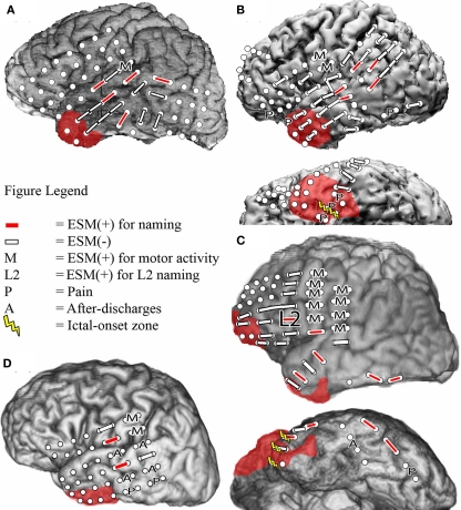
Multilingual patients pose a unique challenge when planning epilepsy surgery near language cortex because the cortical representations of each language may be distinct. These distinctions may not be evident with routine electrocortical stimulation mapping (ESM). Electrocorticography (ECoG) has recently been used to detect task-related spectral perturbations associated with functional brain activation. We hypothesized that using broadband high gamma augmentation (HGA, 60-150 Hz) as an index of cortical activation, ECoG would complement ESM in discriminating the cortical representations of first (L1) and second (L2) languages. We studied four adult patients for whom English was a second language, in whom subdural electrodes (a total of 358) were implanted to guide epilepsy surgery. Patients underwent ECoG recordings and ESM while performing the same visual object naming task in L1 and L2. In three of four patients, ECoG found sites activated during naming in one language but not the other. These language-specific sites were not identified using ESM. In addition, ECoG HGA was observed at more sites during L2 versus L1 naming in two patients, suggesting that L2 processing required additional cortical resources compared to L1 processing in these individuals. Post-operative language deficits were identified in three patients (one in L2 only). These deficits were predicted by ECoG spectral mapping but not by ESM. These results suggest that pre-surgical mapping should include evaluation of all utilized languages to avoid post-operative functional deficits. Finally, this study suggests that ECoG spectral mapping may potentially complement the results of ESM of language.

Keywords: ECoG; electrocortical stimulation mapping; electrocorticography; epilepsy surgery; functional mapping; high gamma; multilingual; naming.


Figures

Figure 1
Figure 1
Bipolar electrocortical stimulation mapping of visual object naming of L1 and L2 in Patient 1 (A), Patient 2 (B), and Patient 3b (C), and only in L2 in Patient 4 (D) using co-registration of a post-surgical CT with pre-surgical MRI to determine electrode placement. Left lateral and left basal views are shown when applicable. White circles indicate the locations of the implanted subdural electrodes. In Patients 1 and 4, the ictal-onset zone was in the left mesial temporal region and is not depicted. Areas with red shading indicate the resection boundaries. No basal temporal ESM was performed in these patients, so basal views are not included.
Figure 2
Figure 2
Electrocorticographic spectral mapping of visual object naming in L1 and L2 in Patient 1 (A), Patient 2 (B), Patient 3a (C), and Patient 4 (D). Lateral, interhemispheric and basal views are shown when applicable. Electrodes used as ground or reference electrodes and those containing electrode artifact are indicated with an “X”. Blue circles indicate HGA(+) electrodes during only L1 visual object naming. Yellow circles indicate HGA(+) electrodes during only L2 visual object naming. Green circles indicate HGA(+) electrodes during both L1 and L2 naming. As in Figure 1, areas with red shading indicate resection boundaries.
Figure 3
Figure 3
Electrocorticographic spectral mapping of visual object naming in L1 and L2 in Patient 3b. Electrode color schemes are the same as in Figure 2. HGA(+) electrodes that were over regions that were ultimately resected are enlarged for emphasis. Time–frequency plots of changes in power obtained with MP analysis are shown for one selected anterior temporal electrode that was HGA(−) for L1 (top) and HGA(+) for L2 (bottom) and was over a region that was ultimately resected. Time zero on the X-axis is the time of stimulus onset. On the color spectrum depicted to the right of each plot, red to yellow indicates a significant increase in magnitude of power after stimulus-onset relative to the baseline within a given time (in ms)–frequency (in Hz) range, and the spectrum from light to dark blue indicates a significant decrease in power. White indicates no significant difference.

Similar articles

Cited by

References

    1. Abutalebi J. (2008). Neural aspects of second language representation and language control. Acta Psychol. (Amst.) 128, 466–478 - PubMed
    1. Aglioti S., Beltramello A., Girardi F., Fabbro F. (1996). Neurolinguistic and follow-up study of an unusual pattern of recovery from bilingual subcortical aphasia. Brain 119, 1551–156410.1093/brain/119.5.1551 - DOI - PubMed
    1. Aglioti S., Fabbro F. (1993). Paradoxical selective recovery in a bilingual aphasic following subcortical lesions. Neuroreport 4, 1359–136210.1097/00001756-199309150-00019 - DOI - PubMed
    1. Alvarez R. P., Holcomb P. J., Grainger J. (2003). Accessing word meaning in two languages: an event-related brain potential study of beginning bilinguals. Brain Lang. 87, 290–30410.1016/S0093-934X(03)00108-1 - DOI - PubMed
    1. Anderson J. M., Gilmore R., Roper S., Crosson B., Bauer R. M., Nadeau S., Beversdorf D. Q., Cibula J., Rogish M., 3rd, Kortencamp S., Hughes J. D., Gonzalez Rothi L. J., Heilman K. M. (1999). Conduction aphasia and the arcuate fasciculus: a reexamination of the Wernicke–Geschwind model. Brain Lang. 70, 1–1210.1006/brln.1999.2135 - DOI - PubMed

LinkOut - more resources