. 2011 Feb 23;6(2):e17001.
doi: 10.1371/journal.pone.0017001.

Clinically translatable cell tracking and quantification by MRI in cartilage repair using superparamagnetic iron oxides

Affiliations

Clinically translatable cell tracking and quantification by MRI in cartilage repair using superparamagnetic iron oxides

Gerben M van Buul et al. PLoS One. .

Abstract

Background: Articular cartilage has very limited intrinsic regenerative capacity, making cell-based therapy a tempting approach for cartilage repair. Cell tracking can be a major step towards unraveling and improving the repair process of these therapies. We studied superparamagnetic iron oxides (SPIO) for labeling human bone marrow-derived mesenchymal stem cells (hBMSCs) regarding effectivity, cell viability, long term metabolic cell activity, chondrogenic differentiation and hBMSC secretion profile. We additionally examined the capacity of synovial cells to endocytose SPIO from dead, labeled cells, together with the use of magnetic resonance imaging (MRI) for intra-articular visualization and quantification of SPIO labeled cells.

Methodology/prinicipal findings: Efficacy and various safety aspects of SPIO cell labeling were determined using appropriate assays. Synovial SPIO re-uptake was investigated in vitro by co-labeling cells with SPIO and green fluorescent protein (GFP). MRI experiments were performed on a clinical 3.0T MRI scanner. Two cell-based cartilage repair techniques were mimicked for evaluating MRI traceability of labeled cells: intra-articular cell injection and cell implantation in cartilage defects. Cells were applied ex vivo or in vitro in an intra-articular environment and immediately scanned. SPIO labeling was effective and did not impair any of the studied safety aspects, including hBMSC secretion profile. SPIO from dead, labeled cells could be taken up by synovial cells. Both injected and implanted SPIO-labeled cells could accurately be visualized by MRI in a clinically relevant sized joint model using clinically applied cell doses. Finally, we quantified the amount of labeled cells seeded in cartilage defects using MR-based relaxometry.

Conclusions: SPIO labeling appears to be safe without influencing cell behavior. SPIO labeled cells can be visualized in an intra-articular environment and quantified when seeded in cartilage defects.


Conflict of interest statement

Competing Interests: The authors have declared that no competing interests exist.

Figures

Figure 1
Figure 1. Labeling human Bone Marrow stroma-derived mesenchymal Stem Cells (hBMSCs) using Superparamagnetic Iron Oxides (SPIO).
Perl's iron stain showing effective endocytosis of SPIO (A), leading to labeling efficiencies of approximately 95% (B) and a total iron load of approximately 20 pg/cell (C). SPIO labeling did not impair cell viability (D) or subsequent metabolic cell activity (E) at any dose used. Results shown for triplicate samples from three hBMSC donors.
Figure 2
Figure 2. Chondrogenic differentiation of hBMSCs is not affected by SPIO labeling.
Collagen II immunohistochemistry and Perl's iron stain demonstrated SPIO labeling to have no effect on pellet size or collagen II production, while iron remained present in differentiated pellets (A–D). Glycosaminoglycan deposition was also unaffected, as can be seen by thionin stain (E–H) and DMB assay (I). Gene expression of collagen II was unaltered by SPIO labeling (J). All analyses were performed after 35 days of differentiation. Insets display low magnification overviews of the entire pellets. Results shown for triplicate samples from two hBMSC donors.
Figure 3
Figure 3. MRI visualization of SPIO-labeled hBMSCs in an intra-articular environment.
Injected cells were visualized in a dose dependent manner (A–D, in-plane resolution 273×273 µm; slice thickness 600 µm). Cells were found scattered throughout the joint (arrows) and could be discerned from intra-articular structures. Labeled cells implanted in cartilage defects in vitro were homogeneously distributed and imaged in a comparable way; dose dependent and clearly distinguishable from structures like cartilage and bone (E–I; in-plane resolution 78×78 µm; slice thickness 1500 µm). After implanting cells in a cartilage defect in an intact porcine knee, MRI showed the cells inside the defect (J; in-plane resolution 273×273 µm; slice thickness 400 µm). In (A); b: bone; c: cartilage; c.l.: cruciate ligament. Intra-articular cells in porcine knees were imaged as a single sample for one donor. hBMSCs implanted in cartilage defects in vitro were imaged in duplicate samples from two hBMSC donors.
Figure 4
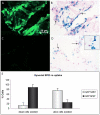
Figure 4. SPIO re-uptake by human synovial explants.
Fluorescent images and Perl's iron stain of GFP-SPIO co-labeled chondrocytes seeded on synovial explants. GFP+-SPIO+ cells, indicating originally seeded chondrocytes, were seen in samples seeded with living cells (A,B). In samples seeded with dead cells, GFP̂-SPIO+ cells were found, indicating SPIO re-uptake by synovial cells (C,D). Furthermore, extracellular SPIO aggregates were observed attached to the synovial explants (D, inset). Approximately 90% of SPIO+ cells in samples seeded with dead chondrocytes were not originally labeled cells, compared with 25% of SPIO+ cells in samples seeded with living cells (E). Arrows indicate SPIO containing cells. Results shown for triplicate samples from two synovium donors.
Figure 5
Figure 5. Quantification of SPIO-labeled cells implanted in cartilage defects in vitro.
Transverse R2* maps from identical osteochondral plugs as shown in figure 3, visualize the differences in labeled cell concentrations (A–E; in-plane resolution 156×156 µm, slice thickness 700 µm). A linear relationship between labeled cell concentration and R2* value was found in a range of 0.4×10⁶–2.6×10⁶ cells/ml (F). Grey voxels inside the defects represent positions where R2* values could not be obtained due to a too low (mainly in A,B) or too high (mainly in E) local concentration of SPIO. Results shown for duplicate samples from two hBMSC donor for samples containing 0; 1.3×10⁵; 4.0×10⁵ and 1.3×10⁶ cells/ml. Samples containing 4.0×10⁴; 8.0×10⁵ and 2.6×10⁶ cells per ml were scanned in duplicates for one hBMSC donor.

Similar articles

Cited by

References

    1. Steinert AF, Ghivizzani SC, Rethwilm A, Tuan RS, Evans CH, et al. Major biological obstacles for persistent cell-based regeneration of articular cartilage. Arthritis Res Ther. 2007;9:213. - PMC - PubMed
    1. Brittberg M, Lindahl A, Nilsson A, Ohlsson C, Isaksson O, et al. Treatment of deep cartilage defects in the knee with autologous chondrocyte transplantation. N Engl J Med. 1994;331:889–895. - PubMed
    1. van Osch GJ, Brittberg M, Dennis JE, Bastiaansen-Jenniskens YM, Erben RG, et al. Cartilage repair: past and future–lessons for regenerative medicine. J Cell Mol Med. 2009;13:792–810. - PMC - PubMed
    1. Centeno CJ, Schultz JR, Cheever M, Robinson B, Freeman M, et al. Safety and complications reporting on the re-implantation of culture-expanded mesenchymal stem cells using autologous platelet lysate technique. Curr Stem Cell Res Ther. 2010;5:81–93. - PubMed
    1. Wakitani S, Nawata M, Tensho K, Okabe T, Machida H, et al. Repair of articular cartilage defects in the patello-femoral joint with autologous bone marrow mesenchymal cell transplantation: three case reports involving nine defects in five knees. J Tissue Eng Regen Med. 2007;1:74–79. - PubMed

Publication types

MeSH terms