. 2011 Jun;54(6):1567-78.
doi: 10.1007/s00125-011-2061-4. Epub 2011 Mar 4.

Cannabinoid 1 receptor activation contributes to vascular inflammation and cell death in a mouse model of diabetic retinopathy and a human retinal cell line

Affiliations

Cannabinoid 1 receptor activation contributes to vascular inflammation and cell death in a mouse model of diabetic retinopathy and a human retinal cell line

A B El-Remessy et al. Diabetologia. 2011 Jun.

Abstract

Aims/hypothesis: Recent studies have demonstrated that cannabinoid-1 (CB(1)) receptor blockade ameliorated inflammation, endothelial and/or cardiac dysfunction, and cell death in models of nephropathy, atherosclerosis and cardiomyopathy. However the role of CB(1) receptor signalling in diabetic retinopathy remains unexplored. Using genetic deletion or pharmacological inhibition of the CB(1) receptor with SR141716 (rimonabant) in a rodent model of diabetic retinopathy or in human primary retinal endothelial cells (HREC) exposed to high glucose, we explored the role of CB(1) receptors in the pathogenesis of diabetic retinopathy.

Methods: Diabetes was induced using streptozotocin in C57BL/6J Cb(1) (also known as Cnr1)(+/+) and Cb(1)(-/-) mice aged 8 to 12 weeks. Samples from mice retina or HREC were used to determine: (1) apoptosis; (2) activity of nuclear factor kappa B, intercellular adhesion molecule 1 (ICAM-1), vascular cell adhesion molecule 1 (VCAM-1), poly (ADP-ribose) polymerase and caspase-3; (3) content of 3-nitrotyrosine and reactive oxygen species; and (4) activation of p38/Jun N-terminal kinase/mitogen-activated protein kinase (MAPK).

Results: Deletion of CB(1) receptor or treatment of diabetic mice with CB(1) receptor antagonist SR141716 prevented retinal cell death. Treatment of diabetic mice or HREC cells exposed to high glucose with SR141716 attenuated the oxidative and nitrative stress, and reduced levels of nuclear factor κB, ICAM-1 and VCAM-1. In addition, SR141716 attenuated the diabetes- or high glucose-induced pro-apoptotic activation of MAPK and retinal vascular cell death.

Conclusions/interpretation: Activation of CB(1) receptors may play an important role in the pathogenesis of diabetic retinopathy by facilitating MAPK activation, oxidative stress and inflammatory signalling. Conversely, CB(1) receptor inhibition may be beneficial in the treatment of this devastating complication of diabetes.


Conflict of interest statement

Duality of interest The authors declare that there is no duality of interest associated with this manuscript.

Figures

Fig. 1
Fig. 1
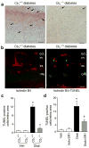
CB1 receptor plays a role in diabetes-induced retinal cell death. a Representative images (×200 magnification, scale bars 25 μm) and (c) statistical analysis showing that diabetes induced significant increases in retinal cell death as indicated by quantitative analysis of TUNEL-positive cells in flat-mounted retina. Deletion of Cb1 completely protected diabetic animals from retinal cell death (n=6). Arrows (a) indicate TUNEL-positive cells in retinal flat mounts. b Co-localisation studies in diabetic retinal sections demonstrated that several TUNEL-positive cells (green) are located within ganglion layer (GCL) and localised with endothelial cells as indicated by isolectin B4 (red); ×200 magnification; scale bars 25 μm. IPL, inner plexiform layer; INL, inner nuclear layer; ONL, outer nuclear layer. d Statistical analysis showing that treatment of diabetic animals (Diab) with SR 141716A (SR1) significantly reduced TUNEL-positive cells compared with diabetic animals treated with vehicle (Veh, n=6). *p<0.05 vs vehicle group; p<0.05 vs wild-type diabetes
Fig. 2
Fig. 2
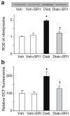
CB1 receptor inhibition attenuates diabetes-induced oxidative and nitrative stress in vivo. a Slot blot and statistical analysis of mouse retinal lysate showing twofold increase in 3-nitrotyrosine formation in diabetic mice as compared with controls (n=5–6). b Statistical analysis showing twofold increase in ROS formation as indicated by DCF fluorescence in diabetic mice as compared with controls (n=6). *p<0.05 vs vehicle group; †p<0.05 vs diabetes. Diab, diabetic animals; SR1, SR 141716A; Veh, vehicle-treated
Fig. 3
Fig. 3
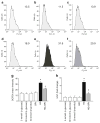
CB1 receptor inhibition attenuates high glucose-induced oxidative stress in HREC. a Representative histograms of DCFDA fluorescence with D-glucose (5 mmol/l), (b) SR 141716A (SR1), (c) L-glucose (30 mmol/l), (d) mannitol (30 mmol/l), (e) high glucose (HG; D-glucose 30 mmol/l) and (f) high glucose + SR1. Mean fluorescence intensity–height at ~525 nm (FL1-H) (a) 13.5, (b) 11.5, (c) 13.9, (d) 16.0, (e) 31.6 and (f) 20.9. g Summary data of DCFDA fluorescence from the indicated representative treatment condition (n=4–6). High glucose (30 mmol/l D-glucose) induced an approximately 2.4-fold increase in DCF fluorescence in HREC cells. h Statistical analysis showing approximately fivefold increase in 3-nitrotyrosine (NT) formation in HREC cells maintained in high glucose as compared with those maintained in normal glucose. Treatment of the cells with CB1 antagonist SR 141716A (2 μmol/l) almost completely prevented high glucose-induced increase in oxidative and nitrative stress. Treatment of HREC with SR1 alone or osmotic controls did not alter ROS or 3-nitrotyrosine formation. *p<0.05 vs vehicle group; p<0.05 vs diabetes
Fig. 4
Fig. 4
CB1 receptor inhibition attenuated diabetes-induced glial activation and NFκB production. a Representative images showing a substantial increase in the intensity of GFAP immunoreactivity in the filaments of Müller cells in diabetes (Diab) vs vehicle (Veh). This increase extended from the nerve fibre layer and inner plexiform layer (IPL) into the outer nuclear layer (ONL) of retina as compared with controls. b Representative images of NFκB in mouse retinal sections showing an approximately 2.5-fold increase in diabetic mice as compared with the controls. NFκB was mainly localised within retinal capillaries (n=6). Magnification (a, b) ×200; scale bars 25 μm. GCL, ganglion cell layer; INL, inner nuclear layer; SR1, SR 141716A. c Statistical analysis of above findings (b); *p<0.05 vs vehicle group; p<0.05 vs diabetes. ROD, relative optic density
Fig. 5
Fig. 5
CB1 receptor inhibition attenuated diabetes-induced adhesion molecule production in vivo. a Representative images of VCAM-1 in mice retinal sections showing an approximately twofold increase in diabetic (Diab) mice as compared with vehicle (Veh)-treated controls. VCAM-1 was mainly localised within retinal capillaries; n=5, ×200 magnification, scale bar 25 μm. GCL, ganglion cell layer; IPL, inner plexiform layer; INL, inner nuclear layer; ONL, outer nuclear layer. c Statistical analysis of above (a) findings, expressed as relative optical density (ROD). b Western blot analysis showing increase in adhesion molecule ICAM-1 levels in diabetic animals as compared with the controls (n=5–6). Treatment of diabetic animals with SR 141716A (SR1) blocked these effects, but did not alter control levels. d Statistical analysis of above findings (b) showing 1.9-fold increase in diabetic animals.*p<0.05 vs vehicle group; p<0.05 vs diabetes
Fig. 6
Fig. 6
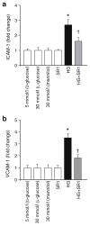
CB1 receptor inhibition attenuated high glucose-induced ICAM-1 and VCAM-1 production in HREC. a Statistical analysis of ICAM-1 and (b) VCAM-1 levels measured by ELISA and showing 2.5- and 3.5-fold increases, respectively in HREC cells maintained in high glucose (HG; 30 mmol/l D-glucose) as compared with those maintained in normal glucose. When the cells were incubated with SR 141716A (SR1; 2 μmol/l), high glucose-induced adhesion molecule production was significantly reduced. Treatment of HREC with SR 141716A alone or osmotic controls did not alter levels of adhesion molecules. *p<0.05 vs vehicle group; p<0.05 vs diabetes
Fig. 7
Fig. 7
CB1 receptor inhibition attenuates diabetes-induced proapoptotic MAPK activation in vivo and in HREC. a Western blot of mice retinal lysate showing an increase in the activation of p38MAPK in diabetic (Diab) as compared with the vehicle (Veh)-treated controls; the increase was mitigated by SR 141716A (SR1) treatment (n=4–6). b Statistical analysis of above results (a), indicating a 2.4-fold increase. c Western blot of HREC cell lysate showing significant increases in activation of p38MAPK and JNK in cells maintained in high glucose (HG; 30 mmol/l D-glucose) as compared with cells maintained in normal glucose. These increases were mitigated upon treatment with SR 141716A (2 μmol/l). d, e Statistical analysis of above (c) findings for MAPK and JNK respectively. *p<0.05 vs vehicle group; p<0.05 vs diabetes
Fig. 8
Fig. 8
CB1 receptor inhibition mitigates high glucose–induced cell death in HREC. a Western blot statistical analysis of HREC cell lysate showing significant increases in the activity of PARP and (b) caspase-3 in cells maintained in high glucose (HG) as compared with cells maintained in normal glucose. These increases were mitigated upon treatment with SR 141716A (SR1; 2 μmol/l). c Flow cytometric analysis of cell death with D-glucose (5 mmol/l), (d) SR 141716A (SR1), (e) L-glucose (30 mmol/l), (f) mannitol (30 mmol/l), (g) high glucose (HG; D-glucose 30 mmol/l) and (h) high glucose + SR1. Cells were maintained in different media as indicated and treated with CB1 antagonists for 1 h, followed by incubation with different media for 48 h in the continuous presence of CB1 antagonists. ch Sytox green (y-axes), x-axes: annexin V-APC. i Summary of the results showing significant increase in apoptosis in HREC cells maintained in high glucose (30 mmol/l D-glucose) compared with those maintained in normal glucose. When the cells were incubated with SR 141716A (2 μmol/l), the high glucose-induced apoptosis was significantly reduced. Treatment of HREC with SR 141716A alone or osmotic controls did not alter cell death. n=4–6; *p<0.05 vs vehicle group; p<0.05 vs diabetes. FL4-H, fluorescence intensity–height at ~675 nm

Similar articles

Cited by

References

    1. Huysman E, Mathieu C. Diabetes and peripheral vascular disease. Acta Chir Belg. 2009;109:587–594. - PubMed
    1. Talahalli R, Zarini S, Sheibani N, Murphy RC, Gubitosi-Klug RA. Increased synthesis of leukotrienes in the mouse model of diabetic retinopathy. Invest Ophthalmol Vis Sci. 2010;51:1699–1708. - PMC - PubMed
    1. Khalfaoui T, Lizard G, Ouertani-Meddeb A. Adhesion molecules (ICAM-1 and VCAM-1) and diabetic retinopathy in type 2 diabetes. J Mol Histol. 2008;39:243–249. - PubMed
    1. Kowluru RA, Kanwar M, Kennedy A. Metabolic memory phenomenon and accumulation of peroxynitrite in retinal capillaries. Exp Diabetes Res. 2007;2007:21976. - PMC - PubMed
    1. El-Remessy AB, Al-Shabrawey M, Khalifa Y, Tsai N-t, Caldwell RB, Liou GI. Neuroprotective and blood–retinal barrier-preserving effects of cannabidiol in experimental diabetes. Am J Pathol. 2006;168:235–244. - PMC - PubMed

Publication types

MeSH terms