. 2011 Jun;461(6):677-86.
doi: 10.1007/s00424-011-0942-5. Epub 2011 Mar 4.

Membrane cholesterol modulates cochlear electromechanics

Affiliations

Membrane cholesterol modulates cochlear electromechanics

William E Brownell et al. Pflugers Arch. 2011 Jun.

Abstract

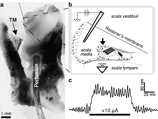
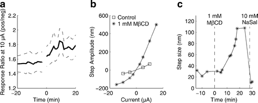
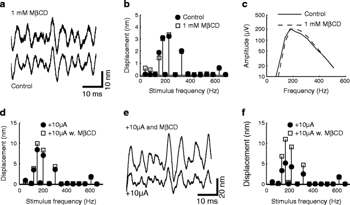
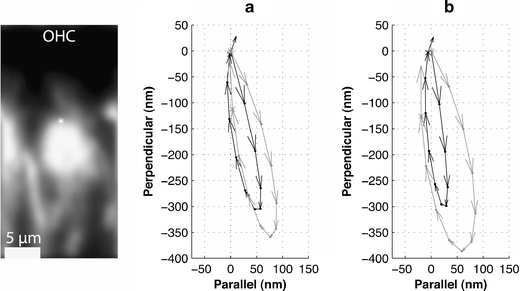
Changing the concentration of cholesterol in the plasma membrane of isolated outer hair cells modulates electromotility and prestin-associated charge movement, suggesting that a similar manipulation would alter cochlear mechanics. We examined cochlear function before and after depletion of membrane cholesterol with methyl-β-cyclodextrin (MβCD) in an excised guinea pig temporal bone preparation. The mechanical response of the cochlear partition to acoustic and/or electrical stimulation was monitored using laser interferometry and time-resolved confocal microscopy. The electromechanical response in untreated preparations was asymmetric with greater displacements in response to positive currents. Exposure to MβCD increased the magnitude and asymmetry of the response, without changing the frequency tuning of sound-evoked mechanical responses or cochlear microphonic potentials. Sodium salicylate reversibly blocked the enhanced electromechanical response in cholesterol depleted preparations. The increase of sound-evoked vibrations during positive current injection was enhanced following MβCD in some preparations. Imaging was used to assess cellular integrity which remained unchanged after several hours of exposure to MβCD in several preparations. The enhanced electromechanical response reflects an increase in outer hair cell electromotility and may reveal features of cholesterol distribution and trafficking in outer hair cells.
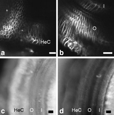

Figures

Fig. 1
Fig. 1
Experimental methods. a Photomicrograph of excised temporal bone preparation. Tympanic membrane (TM) and malleus are visible on the left. A perfusion tube is inserted through an opening into the scala tympani in the basal turn of the cochlea and oxygenated artificial perilymph is introduced under ~10-cm pressure. The endpiece of the tubing has a smaller diameter and the hole in the cochlea is smaller than the visible tubing. Perfusion solutions exit through an opening in the apical turn of the cochlea (visible near the cochlear apex). Optical recordings of cochlear movements are made and a current stimulus/CM recording micropipette is inserted into the scala media through the apical opening. A tube from the earphone is connected to a rubber O-ring cemented to the remains of the external auditory meatus (not shown). The entire preparation is submerged in artificial perilymph. b Schematic of a radial section of the apical cochlea showing the relative position of the micropipette and the organ of Corti. Laser (arrowhead) for interferometry is focused on a highly reflective Hensen’s cell lipid droplet. High-speed confocal imaging and time-resolved measurements are also made through a similar opening. c A single trace of electromechanically evoked displacement riding on top of smaller acoustically evoked vibrations
Fig. 2
Fig. 2
Electromechanical response of the apical cochlear partition. a–c Response to positive and d–f negative 10-μA current injections. Control responses (a, d) and the response from the same preparation after perfusion with 1 mM MβCD (b, e) from a representative preparation. The responses are fit to single exponential functions (thin solid lines). c, f Summary data for nine preparations showing the evolution of the normalized response to ±10-μA current injections. Summary responses are normalized to the control response 20 min prior to perfusion. The dashed lines show standard error of the mean at each time interval. Values at zero time have been blanked out because the mean and S.E.M. were fit with a 5-point sliding interpolation function which introduced artifactual values at zero time. The top summary plot panel also shows the evolution of the normalized response to positive currents in six control preparations. The values were collected for 20 min before and after 1.5 h of cochlear perfusion (MβCD was typically perfused between 1 and 1.5 h of initiating cochlear perfusion)
Fig. 3
Fig. 3
The magnitude of the response to positive current is greater than the response to negative current and salicylate blocks the electromechanical response. a Summary plot of the ratio of the magnitude of the response to positive current to the magnitude of the negative current for nine preparations. b Response of a representative preparation to current steps of different magnitude before and after perfusion with MβCD. c Electromechanical responses over time from a representative preparation to +10-μA current injections showing typical increase in response to MβCD perfusion followed by a rapid decline in response to perfusion with 10 mM NaSal
Fig. 4
Fig. 4
Cochlear partition tuning and cochlear microphonics are similar before and after exposure to MβCD. a Time domain and b spectral analysis of sound-evoked mechanical response. c Cochlear microphonics from the same preparation as a and b are similar before and after MβCD. d Spectra from another preparation showing a modest increase in sound-evoked response during a +10-μA current injection following exposure to MβCD. e Time domain and f spectral analysis of sound-evoked response during a +10-μA current injection from a preparation showing greater response following MβCD
Fig. 5
Fig. 5
Confocal imaging of sound-evoked vibrations confirms a larger effect of positive current injection following MβCD. Confocal image on the left of outer hair cell recorded in situ during simultaneous sound and electrical stimulation. Vibrations at the base of the stereocilia bundle were measured at a point on the reticular lamina marked by the dot. a, b Acoustically evoked vibration trajectories during negative (dark arrows) and positive (gray arrows) stimulation before (a) and after (b) exposure to MβCD. Movements parallel to the reticular lamina are plotted along the abscissa while perpendicular movements are plotted along the ordinate
Fig. 6
Fig. 6
Cochlear histology following MβCD exposure. a, b Confocal images of the cochlea after 10 min of perfusion with RH795. a Optical section through Reissner’s membrane (RM) and Hensen’s cells (HeC) near the site of vibration measurements. b Optical section through the organ of Corti at a deeper focal plane than a. (I, inner hair cells; O, outer hair cells). c UV fluorescence microscopy of filipin-labeled organ of Corti in a control preparation not exposed to MβCD. d Filipin labeling in a preparation perfused with 1 mM MβCD. All scale bars = 25 μm

References

    1. Abe T, Kakehata S, Kitani R, Maruya S, Navaratnam D, Santos-Sacchi J, Shinkawa H. Developmental expression of the outer hair cell motor prestin in the mouse. J Membr Biol. 2007;215:49–56. doi: 10.1007/s00232-007-9004-5. - DOI - PMC - PubMed
    1. Adler HJ, Belyantseva IA, Merritt RC, Jr, Frolenkov GI, Dougherty GW, Kachar B. Expression of prestin, a membrane motor protein, in the mammalian auditory and vestibular periphery. Hear Res. 2003;184:27–40. doi: 10.1016/S0378-5955(03)00192-8. - DOI - PubMed
    1. Alberts B. Molecular biology of the cell. 4. New York: Garland Science; 2002.
    1. Ashmore J, Avan P, Brownell WE, Dallos P, Dierkes K, Fettiplace R, Grosh K, Hackney CM, Hudspeth AJ, Julicher F, Lindner B, Martin P, Meaud J, Petit C, Sacchi JR, Canlon B. The remarkable cochlear amplifier. Hear Res. 2010;266:1–17. doi: 10.1016/j.heares.2010.05.001. - DOI - PMC - PubMed
    1. Belyantseva IA, Adler HJ, Curi R, Frolenkov GI, Kachar B. Expression and localization of prestin and the sugar transporter GLUT-5 during development of electromotility in cochlear outer hair cells. J Neurosci. 2000;20:116. - PMC - PubMed

Publication types