Case Reports
. 2011 Jun;258(6):1098-104.
doi: 10.1007/s00415-010-5892-x. Epub 2011 Mar 4.

SMART syndrome: a late reversible complication after radiation therapy for brain tumours

Affiliations
Case Reports

SMART syndrome: a late reversible complication after radiation therapy for brain tumours

Joost P Kerklaan et al. J Neurol. 2011 Jun.

Abstract

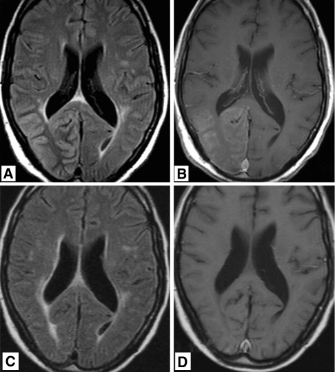
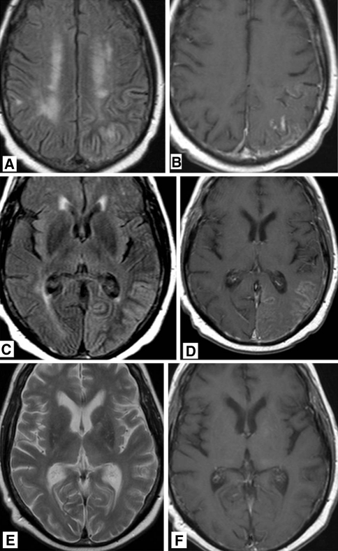
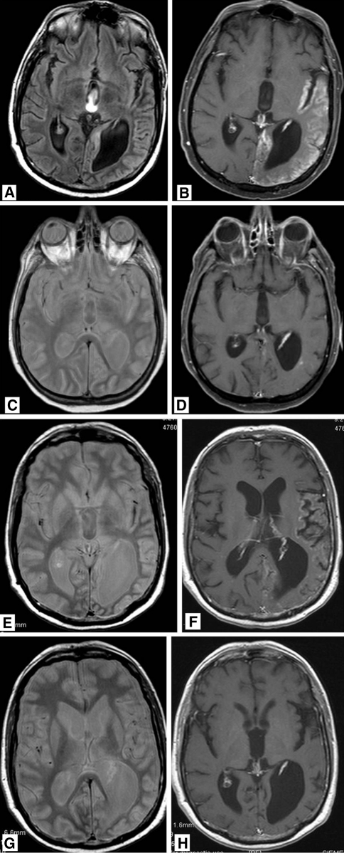
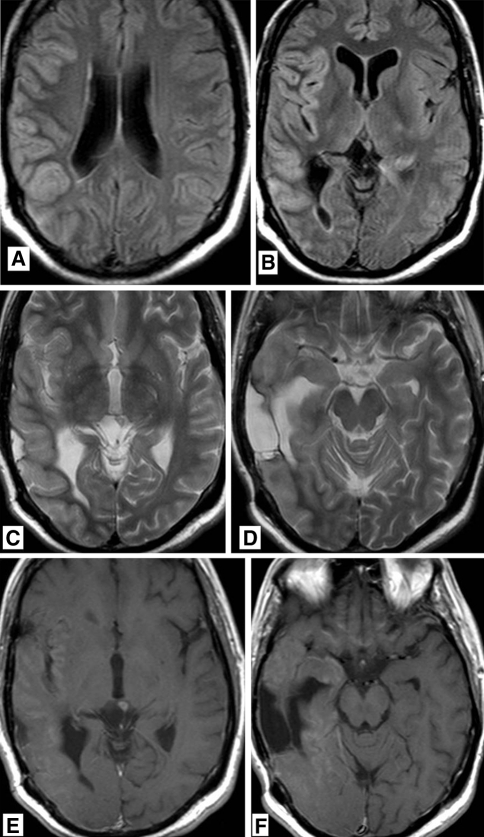
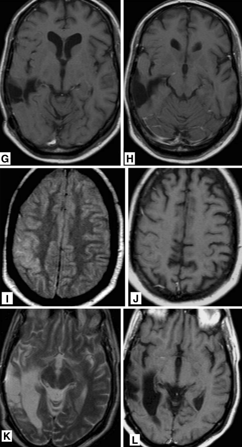
With intensified treatment leading to longer survival, complications of therapy for brain tumours are more frequently observed. Regarding radiation therapy, progressive and irreversible white matter disease with cognitive decline is most feared. We report on four patients with reversible clinical and radiological features occurring years after radiation for brain tumours, suggestive for the so called SMART syndrome (stroke-like migraine attacks after radiation therapy). All four patients (males, age 36-60 years) had been treated with focal brain radiation for a primary brain tumour or with whole-brain radiation therapy for brain metastases. Ranging from 2 to 10 years following radiation therapy patients presented with headache and focal neurological deficits, suggestive for tumour recurrence. Two patients also presented with focal seizures. MRI demonstrated typical cortical swelling and contrast enhancement, primarily in the parieto-occipital region. On follow-up both clinical and MRI features improved spontaneously. Three patients eventually proved to have tumour recurrence. The clinical and radiological picture of these patients is compatible with the SMART syndrome, a rare complication of radiation therapy which is probably under recognized in brain tumour patients. The pathophysiology of the SMART syndrome is poorly understood but bears similarities with the posterior reversible encephalopathy syndrome (PRES). These four cases underline that the SMART syndrome should be considered in patients formerly treated with radiation therapy for brain tumours, who present with new neurologic deficits. Before the diagnosis of SMART syndrome can be established other causes, such as local tumour recurrence, leptomeningeal disease or ischemic disease should be ruled out.


Figures

Fig. 1
Fig. 1
36-year-old man who was treated 4 years previously for pineal tumor with radiation therapy, now presenting with headache and left hemianopia. a T2 weighted, fluid attenuated inversion recovery (FLAIR) MR image showing diffuse gyral swelling and signal increase in the right occipital lobe. b T1 weighted MR image after gadolinium administration showing diffuse gyral and leptomeningeal enhancement. MR images obtained 9 month later showing non residual signal abnormalities on FLAIR (c) and no abnormal enhancement (d)
Fig. 2
Fig. 2
60-year-old man who was treated 10 years previously for brain metastases of bronchial carcinoma with chemo- and radiation therapy, now presenting with gradually progressive headache and acute dysphasis. a T2 weighted MR image showing diffuse gyral swelling and signal increase in the left parietal lobe. b T1 weighted MR image after gadolinium administration showing abnormal regional leptomeningeal enhancement. c T2 weighted FLAIR MR image showing diffuse gyral swelling and signal increase in the left occipital lobe. d T1 weighted MR image after gadolinium administration showing abnormal leptomeningeal enhancement. MR images after 4 months. e T2 weighted MR image no residual signal abnormalities. f T1 weighted MR image after gadolinium administration showing no abnormal leptomeningeal enhancement
Fig. 3
Fig. 3
47-year-old man who was treated 5 years previously for a left parietal oligodendrolioma with chemo- and radiation therapy, now presenting with right hemianopia and mild paresis of his right arm and leg. T2 weighted FLAIR MR images a showing gyral swelling and signal increase in the temporal lobe. b After gadolinium administration, diffuse gyral enhancement is seen in the occipital, temporal and insular lobe. Proton density (c) and T1 post gadolinium (d) MR imaging 8 days later showing normalization of signal intensity in the temporal lobe and marked reduction in abnormal gyral enhancement. After 3 years, the patient presented again with right sided paresis. MR imaging again shows signal increase in the temporal lobe on PD weighted images (e) while on T1 post gadolinium imaging (f) abnormal gyral enhancement is again visible. Like after the previous episode, the MR abnormalities disappeared (g, h)
Fig. 4
Fig. 4
38-year-old man who was treated 5 years previously for a right temporal anaplastic astrocytoma with chemo- and radiation therapy, now presenting with increasing epileptic activity and gradually progressive left sided hemiparesis and dysarthria. T2 weighted FLAIR MR images (a, b) showing gyral swelling and signal increase in the temporal lobe. T2 weighted MR image (c, d) showing gyral swelling and signal increase in the temporal lobe. A post-operative parenchymal defect is seen in the area of the previously treated astrocytoma. T1 weighted MR images after gadolinium administration (e, f) abnormal gyral and leptomeningeal enhancement in the temporal lobe and in the basal frontal areas. No focal enhancement in post-operative parenchymal defect. MR images 3 months later. g, h T1 weighted MR image after gadolinium administration showing persistent gyral abnormalities but disappearance of abnormal enhancement. MR images 6 months after treatment for recurrent anaplastic astrocytoma using chemotherapy. i T2 weighted FLAIR MR image and j T1 weighted MR image after gadolinium administration showing persistent gyral abnormalities but disappearance of abnormal enhancement. MR images 6 months after treatment for recurrent anaplastic astrocytoma using chemotherapy. k T2 weighted MR image and l T1 weighted MR image after gadolinium administration resolution of T2 abnormalities and disappearance of abnormal enhancement in the deep temporal area
Fig. 4
Fig. 4
38-year-old man who was treated 5 years previously for a right temporal anaplastic astrocytoma with chemo- and radiation therapy, now presenting with increasing epileptic activity and gradually progressive left sided hemiparesis and dysarthria. T2 weighted FLAIR MR images (a, b) showing gyral swelling and signal increase in the temporal lobe. T2 weighted MR image (c, d) showing gyral swelling and signal increase in the temporal lobe. A post-operative parenchymal defect is seen in the area of the previously treated astrocytoma. T1 weighted MR images after gadolinium administration (e, f) abnormal gyral and leptomeningeal enhancement in the temporal lobe and in the basal frontal areas. No focal enhancement in post-operative parenchymal defect. MR images 3 months later. g, h T1 weighted MR image after gadolinium administration showing persistent gyral abnormalities but disappearance of abnormal enhancement. MR images 6 months after treatment for recurrent anaplastic astrocytoma using chemotherapy. i T2 weighted FLAIR MR image and j T1 weighted MR image after gadolinium administration showing persistent gyral abnormalities but disappearance of abnormal enhancement. MR images 6 months after treatment for recurrent anaplastic astrocytoma using chemotherapy. k T2 weighted MR image and l T1 weighted MR image after gadolinium administration resolution of T2 abnormalities and disappearance of abnormal enhancement in the deep temporal area

References

    1. Kim JH, Brown SL. Mechanisms of radiation-induced brain toxicity and implications for future clinical trials. J Neurooncol. 2008;87:279–286. doi: 10.1007/s11060-008-9520-x. - DOI - PubMed
    1. Lachance DH, Black DF (2005) SMART: strokelike migraine attacks after radiation therapy. Neurology (Suppl 1) A220
    1. Pruitt A, Dalmau J, Detre J. Episodic neurologic dysfunction with migraine and reversible imaging findings after radiation. Neurology. 2006;67:676–678. doi: 10.1212/01.wnl.0000228862.76269.62. - DOI - PubMed
    1. Black DF, Bartleson JD, Bell ML, Lachance DH. Smart: stroke-like migraine attacks after radiation therapy. Cephalagia. 2006;26:1137–1142. doi: 10.1111/j.1468-2982.2006.01184.x. - DOI - PubMed
    1. Bartleson JD, Krecke KN. Reversible, stroke like migraine attacks in patients with previous radiation therapy. Neuro-oncology. 2003;5:121–127. - PMC - PubMed

Publication types