. 2011 Apr;28(4):351-66.
doi: 10.1007/s10585-011-9373-0. Epub 2011 Mar 4.

Loss of one Tgfbr2 allele in fibroblasts promotes metastasis in MMTV: polyoma middle T transgenic and transplant mouse models of mammary tumor progression

Affiliations

Loss of one Tgfbr2 allele in fibroblasts promotes metastasis in MMTV: polyoma middle T transgenic and transplant mouse models of mammary tumor progression

Wei Bin Fang et al. Clin Exp Metastasis. 2011 Apr.

Abstract

Accumulation of fibroblasts is a phenomenon that significantly correlates with formation of aggressive cancers. While studies have shown that the TGF-β signaling pathway is an important regulator of fibroblast activation, the functional contribution of TGF-β signaling in fibroblasts during multi-step tumor progression remains largely unclear. In previous studies, we used a sub-renal capsule transplantation model to demonstrate that homozygous knockout of the Tgfbr2 gene (Tgbr2(FspKO)) enhanced mammary tumor growth and metastasis. Here, we show for the first time a significant role for loss of one Tgfbr2 allele during multi-step mammary tumor progression. Heterozygous deletion of Tgfbr2 in stromal cells in MMTV-PyVmT transgenic mice (PyVmT/Tgfbr2(hetFspKO) mice) resulted in earlier tumor formation and increased stromal cell accumulation. In contrast to previous studies of Tgbr2(FspKO) fibroblasts, Tgfbr2(hetFspKO) fibroblasts did not significantly increase tumor growth, but enhanced lung metastasis in PyVmT transgenic mice and in co-transplantation studies with PyVmT mammary carcinoma cells. Furthermore, Tgfbr2(hetFspKO) fibroblasts enhanced mammary carcinoma cell invasiveness associated with expression of inflammatory cytokines including CXCL12 and CCL2. Analyses of Tgbr2(FspKO) and Tgfbr2(hetFspKO) fibroblasts revealed differences in the expression of factors associated with metastatic spread, indicating potential differences in the mechanism of action between homozygous and heterozygous deletion of Tgfbr2 in stromal cells. In summary, these studies demonstrate for the first time that loss of one Tgfbr2 allele in fibroblasts enhances mammary metastases in a multi-step model of tumor progression, and demonstrate the importance of clarifying the functional contribution of genetic alterations in stromal cells in breast cancer progression.


Figures

Fig. 1
Fig. 1
Loss of one Tgfbr2 allele enhances mammary tumor progression in PyVmT transgenic mice. a PyVmT controls (n = 38) and PyVmT/Tgfbr2hetFspKO (n = 24) mice were palpated for tumors and scored for onset of primary tumor growth. b Tumor burden was assessed by a measurement of total tumor volume at 13.4 weeks of age for control mice and PyVmT/Tgfbr2hetFspKO mice. c H&E stain of primary mammary tumors. The areas outlined with dotted lines indicate necrosis. d Lung metastases were visualized by whole mount hematoxylin staining of lung tissue extracted from PyVmT controls or PyVmT/Tgfbr2hetFspKO mice and scored. Each point in the graph represents the number of lung metastases in one mouse. Bars represent the mean? standard error of the mean (SEM). The presence of nodules was confirmed by H&E stain of 5 micron sections. Representative nodule in PyVmT control is outlined by dotted line. Statistical analysis of PyVmT/Tgfbr2hetFspKO compared to PyVmT controls was performed using two tailed Student’s t-test. Statistical significance determined by P value less than 0.05. *p < 0.01,**p >0.05, ***p < 0.05. Scale bar = 80 μm
Fig. 2
Fig. 2
Primary tumors in PyVmT/Tgfbr2hetFspKO mice show increased tumor cell proliferation and survival. a Ki67 immunostaining as a measure of tumor cell proliferation. b TUNEL immunostaining as a measure of cellular apoptosis. Five random sections were photographed and quantitated for positive staining. PyVmT/Tgfbr2het-FspKO measurements were compared to PyVmT controls using two tailed Student’s t-test. Statistical significance determined by P-value less than 0.05. *p < 0.05, **p < 0.02. Scale bar of low magnified image = 150 μm. Smaller boxed areas within each image refer to magnified inset. Scale bar of inset = 15 μm. Images are representative of the entire area of each the five random sections counted
Fig. 3
Fig. 3
PyVmT/Tgfbr2hetFspKO primary tumors exhibit increased stromal cell accumulation. Tumor sections of PyVmT controls and PyVmT/Tgfbr2hetFspKO were stained with antibodies. a Alpha smooth muscle actin (α-sma), b Fsp1, c vimentin, d F4/80, e Von Willibrand Factor 8. Five random sections were photographed and quantitated for positive staining. Positive DAB staining is emphasized by arrowhead. Statistical analysis of PyVmT/Tgfbr2hetFspKO mice compared to PyVmT controls was performed using Poisson Regression. Statistical significance determined by P-value less than 0.05. *p < 0.04, **p < 0.01, ***P = 05. Scale bar of low magnified image = 150 μm. Small boxed areas within each image refer to magnified inset. Scale bar of inset = 15 μm. Images are representative of the entire area of each the five random sections counted
Fig. 4
Fig. 4
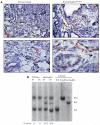
Loss of one Tgfbr2 allele occurs in a subset of mammary fibroblasts from PyVmT/Tgfbr2hetFspKO mice. A Cre immunostaining of primary tumor sections at low (a, b) and high magnification (c, d). Arrows indicate Cre staining in mammary stromal cells. (a, b; bar = 40 μm. c, d; bar = 10 μm). B Southern blot analysis of mammary fibroblasts isolated from control PyVmT or PyVmT/Tgfbr2hetFspKO tumors. Controls include: WT Wildtype fibroblasts, Flox/Flox Fibroblasts carrying homozygous Floxed Tgfbr2, FspKO Homozygous Tgfbr2 knockout fibroblasts. Percent recombination (% recom) was determined by Phosphoimager analysis. Flox Floxed Tgfbr2, KO Knockout of the Tgfbr2 allele by excision of exon 2, WT Wildtype Tgfbr2
Fig. 5
Fig. 5
Loss of one Tgfbr2 allele in mammary fibroblasts enhances overall fibroblast proliferation but does not significantly inhibit cell proliferation in response to TGF-β treatment. a Heterogeneous or b clonal fibroblasts were treated with 5 ng/ml TGF-β and analyzed for changes in cell proliferation by 3[H] thymidine incorporation. Statistical analysis was performed using Two-Way ANOVA, accompanied by post-hoc pair-wise analysis using Bonferroni–Dunn test. Statistical significance determined by P-value less than 0.05. *(–) TGF-β versus (+)TGF-β treatment in a given sample; P < 0.05. **WT/Flox #1 versus WT/Flox #2; P > 0.05. ***hetFspKO #1 versus hetFspKO #2; P > 0.05. ****hetFspKO #1 or #2 versus WT/Flox #1 or #2; P < 0.01
Fig. 6
Fig. 6
Effect of Tgfbr2hetFspKO fibroblasts on PyVmT carcinoma cell invasion. PyVmT carcinoma cells labeled with CMFDA CellTracker dye were embedded in collagen with clonal Tgfbr2Wt/Flox or Tgfbr2hetFspKO fibroblasts and analyzed for invasion through collagen for 72 h. Representative images of PyVmT cells embedded in collagen alone for 48 h are shown. The border of the collagen through which tumor cells invade is indicated by arrow in the magnified images on the right. Experiments were performed in triplicate, n = 3 per experimental group. Statistical analysis of was performed using two-tailed Student’s t-test. Statistical significance determined by P-value less than 0.05. *p < 0.05 versus WT/Flox
Fig. 7
Fig. 7
Tgfbr2hetFspKO fibroblasts enhanced mammary carcinoma invasion through CCL2 but not HGF dependent mechanisms. Conditioned medium was analyzed by ELISA for levels of a HGF, b CCL2 from heterogeneous fibroblasts and c CCL2 from clonal fibroblasts. d PyVmT cells were embedded in collagen with Tgfbr2hetFspKO fibroblasts in the presence or absence of control IgG, anti-CCL2 for 72 h and analyzed for degree of invasion through the collagen. Experiments were performed in triplicate, n = 3 per experimental group. Statistical analysis of was performed using two-tailed Student’s t-test. Statistical significance determined by P-value less than 0.05. *p >0.05 versus WT/Flox, **p < 0.05 versus WT/Flox, ***p < 0.01 versus WT/Flox, ****p < 0.05 versus Rat IgG

Similar articles

Cited by

References

    1. Barrett-Lee P, Travers M, Luqmani Y, et al. Transcripts for transforming growth factors in human breast cancer: clinical correlates. Br J Cancer. 1990;61(4):612–617. - PMC - PubMed
    1. Desmouliere A, Guyot C, Gabbiani G. The stroma reaction myofibroblast: a key player in the control of tumor cell behavior. Int J Dev Biol. 2004;48(5-6):509–517. - PubMed
    1. Kalluri R, Zeisberg M. Fibroblasts in cancer. Nat Rev Cancer. 2006;6(5):392–401. - PubMed
    1. Camps J, Chang S, Hsu T, et al. Fibroblast-mediated acceleration of human epithelial tumor growth in vivo. Proc Natl Acad Sci USA. 1990;87:75–79. - PMC - PubMed
    1. Sakakura T, Sakagami Y, Nishizuka Y. Accelerated mammary cancer development by fetal salivary mesenchyma isografted to adult mouse mammary epithelium. J Natl Cancer Inst. 1981;66(5):953–959. - PubMed

Publication types

MeSH terms

Substances

LinkOut - more resources