. 2011 May;80(3):835-52.
doi: 10.1111/j.1365-2958.2011.07615.x. Epub 2011 Mar 22.

DsbA2 (27 kDa Com1-like protein) of Legionella pneumophila catalyses extracytoplasmic disulphide-bond formation in proteins including the Dot/Icm type IV secretion system

Affiliations

DsbA2 (27 kDa Com1-like protein) of Legionella pneumophila catalyses extracytoplasmic disulphide-bond formation in proteins including the Dot/Icm type IV secretion system

Max Jameson-Lee et al. Mol Microbiol. 2011 May.

Abstract

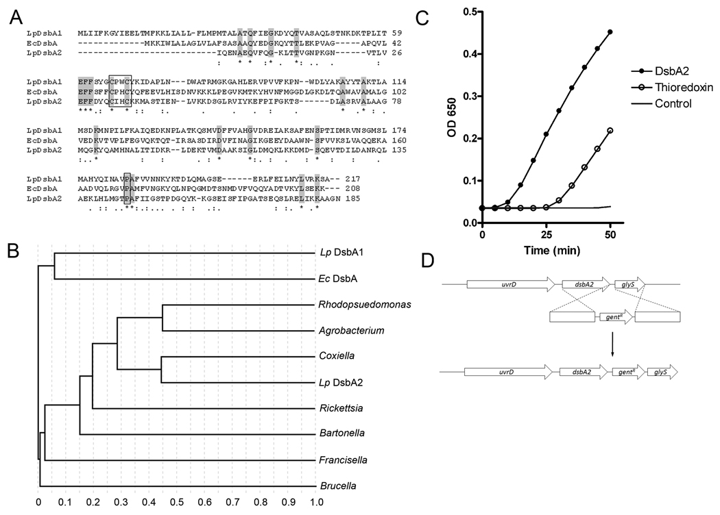
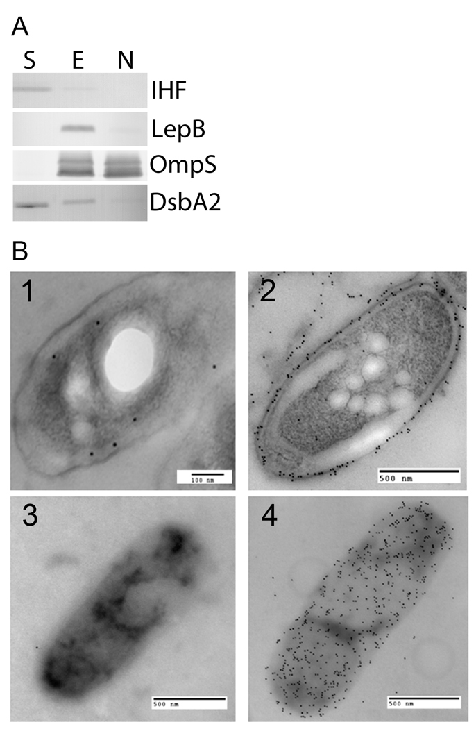
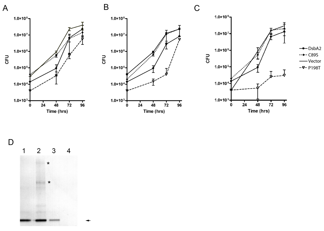
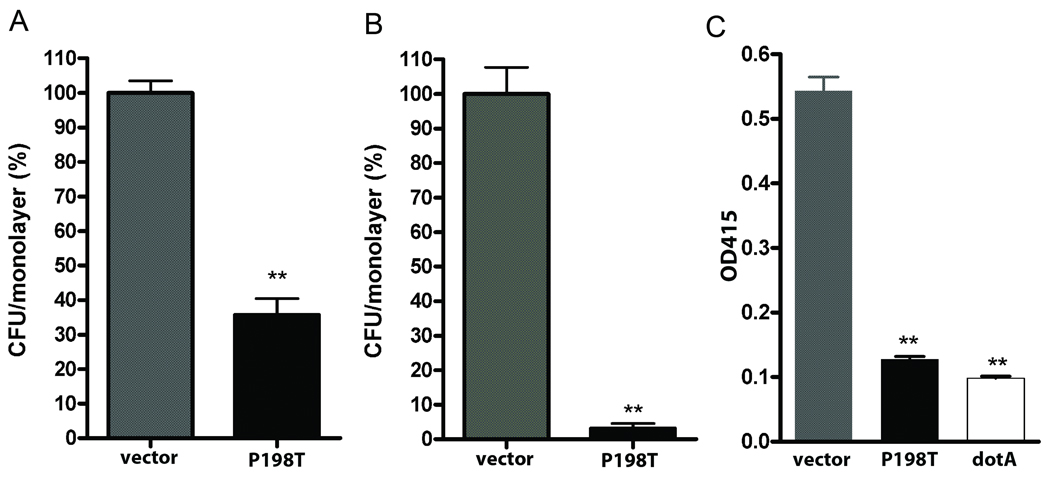
In Gram-negative bacteria, thiol oxidoreductases catalyse the formation of disulphide bonds (DSB) in extracytoplasmic proteins. In this study, we sought to identify DSB-forming proteins required for assembly of macromolecular structures in Legionella pneumophila. Here we describe two DSB-forming proteins, one annotated as dsbA1 and the other annotated as a 27 kDa outer membrane protein similar to Com1 of Coxiella burnetii, which we designate as dsbA2. Both proteins are predicted to be periplasmic, and while dsbA1 mutants were readily isolated and without phenotype, dsbA2 mutants were not obtained. To advance studies of DsbA2, a cis-proline residue at position 198 was replaced with threonine that enables formation of stable disulphide-bond complexes with substrate proteins. Expression of DsbA2 P198T mutant protein from an inducible promoter produced dominant-negative effects on DsbA2 function that resulted in loss of infectivity for amoeba and HeLa cells and loss of Dot/Icm T4SS-mediated contact haemolysis of erythrocytes. Analysis of captured DsbA2 P198T-substrate complexes from L. pneumophila by mass spectrometry identified periplasmic and outer membrane proteins that included components of the Dot/Icm T4SS. More broadly, our studies establish a DSB oxidoreductase function for the Com1 lineage of DsbA2-like proteins which appear to be conserved among those bacteria also expressing T4SS.


Figures

Fig. 1
Fig. 1. Phylogenetic analysis and thioredoxin activity of DsbA2 (Lpg1841)
(A) Amino acid sequence alignments: L. pneumophila AAU26230.1 (Lp DsbA), E. coli CAA56736.1 (Ec DsbA), and L. pneumophila YP_095867.1 (Lp DsbA2). Conserved amino acids are highlighted; active site CXXC and conserved cis-proline residues are boxed. (B) ClustalW phylogenetic analysis delineates a sub-set of DsbA-like oxidoreductases present in several T4SS containing bacteria. Strains listed in Fig. 1A above and Rhodopseudomonas palustris NP_946373.1 (Rhodopseudomonas), Agrobacterium tumafaciens AAK87124.1 (Agrobacterium), Coxiella burnetti BAA20499.1 (Coxiella), Rickettsia typhi YP_067376.1 (Rickettsia), Bartonella henselae YP_033613.1 (Bartonella), Francisella tularensis YP_170079.1 (Francisella), and Brucella melitensis NP_540478.1 (Brucella). All proteins contain CxxC active site and conserved cis-proline. (C) Precipitation of insulin was followed spectrophotometrically after the addition of 1 µM thioredoxin (open circles), 5 µM DsbA2 (dark circles), or no enzyme, DTT control (no symbol). A representative experiment is shown. (D) Genetic organization of the dsbA2 locus and allelic replacement strategy to introduce a gentamicin cassette downstream of dsbA2. Insertions within dsbA2 were not obtained and considered lethal.
Fig. 2
Fig. 2. DsbA2 is not an outer membrane protein
(A) DsbA2 partitions into the soluble phase after Triton X-100 separation. Whole-cell lysates of L. pneumophila were cleared by low-speed centrifugation (10,000 × g), and membrane material was pelleted by ultra-centrifugation (100,000 × g). The Triton-extractable inner membrane proteins (Lane E) were re-suspended in 1% Triton X-100, and the Triton-insoluble outer membrane proteins (Lane N) were re-pelleted by ultra-centrifugation. Equivalent protein amounts were loaded in all lanes, and to ensure fraction separation, a known soluble phase protein (IHF), inner membrane protein (LepB), and outer membrane protein (OmpS) were included as controls for the soluble (S), inner membrane (E), and outer membrane protein (N) fractions, respectively. (B) Immunogold electron microscopy of ultrathin sections and whole cells of L. pneumophila. Panels 1 and 3 are developed with DsbA2 primary antibody and 10 nm gold conjugated goat anti-mouse IgG secondary antibodies, while Panels 2 and 4 are developed with OmpS-specific antibody and 10 nm gold conjugated goat anti-rabbit antibodies. Counting procedures (detailed in the Methods) indicate that DsbA2 is located in the periplasm 63% of the time with 95% confidence. The DsbA2-specific antibody does not cross react with DsbA1.
Fig. 3
Fig. 3. Expression of P198T mutant protein inhibits intracellular growth in amoeba
Acanthamoeba castellanii were infected with L. pneumophila AA100 constructs (A) in the absence of IPTG induction, (B) induction at 0.5 mM IPTG, and (C) at 5 mM IPTG. Colony forming units (cfu) were determined in triplicate at the indicated times in a single experiment: AA100 pMMB:empty (no symbol), DsbA2 (filled circle), DsbA2 C89S (dashed line), or DsbA2 P198T (open triangle). (D) Immunoblot detection of DsbA2 proteins expressed during infection of A. castellenii. Lanes 1. pMMB: DsbA2; Lane 2, pMMB:C89S; Lane 3, pMMB:empty; and Lane 4, pMMB:P198T. DsbA2 at 30 kDa is indicated by the arrow. SDS PAGE was run under non-reducing conditions and higher molecular weight complexes than DsbA2 can be seen in bacteria expressing wild type DsbA2 (Lane 1) and C89S (Lane 3) that disappear under reducing conditions (data not shown). Asterisks denote bands unique to the C89S mutant.
Fig. 4
Fig. 4. Expression of P198T mutant protein inhibits infectivity of HeLa cells and contact lysis of erythrocytes
Effects of P198T expression on attachment to and invasion of HeLa cell monolayers. (A) Attachment of AA100 expressing P198T (black) to HeLa cells was reduced by 60% compared with the empty vector control (grey). (B) Invasion of HeLa cells (gentamicin treatment) by AA100 P198T (black) was decreased by >90% compared to the empty vector control (grey). (C) Erythrocyte contact lysis assay. Erythrocyte lysis was measured at A450 with the AA100 (empty vector) control (grey), AA100 expressing P198T (black) and a dotA mutant that is defective in T4SS (white). ** denotes statistical significance of < 0.05 by student t-test.
Fig. 5
Fig. 5. Expression of P198T mutant protein inhibits flagellar synthesis
Coomassie stained SDS PAGE of the osmotic shockate from the AA100 P198T mutant strain after induction with 1 mM IPTG (+) or uninduced (−). The 55 kDa band noted by the arrow in the uninduced lane and absent from the induced lane was identified as flagellin by MALDI TOF MS. * indicate periplasmic proteins that are identical between induced and uninduced conditions. Lane M denotes molecular weight markers in kilodaltons (kD). WB is a western blot with DsbA2-specific antibody indicating that DsbA2 is present in the shockate.
Fig. 6
Fig. 6. DsbA2 P198T inhibits DsbA2 activity in a modified insulin reduction assay
(A) DsbA2 P198T (5 µM, open triangle) has no activity greater than control (solid line) in the insulin reduction assay. DsbA2 activity in this assay (solid circle) is not affected by addition of 25 µM BSA (star). (B) Modified insulin reduction assay (see text): Insulin precipitation by 5 µM DsbA2 (black circle) and competition with 5 µM (dark dashed triangles) or 25 µM P198T (dark dashed diamonds) mutant protein. As noted in panel A, P198T (open triangle) displayed no activity over that of the DTT control (solid line). The dose dependent effect of the P198T mutant protein on DsbA2 activity is consistent with the possibility that the mutant protein, by binding insulin, has a rate limiting effect on the activity of DsbA2. These experiments were repeated three separate times, representative experiment shown.
Fig. 7
Fig. 7. Substrate-protein capture by P198T-mutant protein in L. pneumophila
(A). Immunoblots (DsbA2 antibodies) of whole cell lysates subjected to SDS-PAGE under non-reducing conditions or as indicated: Lane 1 MW markers; Lane 2, AA100 pMMB:P198T un-induced whole cell lysates; Lane 3 AA100 pMMB:P198T induced with 1 mM IPTG; Lane 4, same as Lane 3 except run under reducing conditions (note disappearance of higher molecular weight complexes). (B) His6 DsbA2-substrate complexes enriched for by Ni++ column chromatography and separated by non-reducing SDS PAGE: Coomassie stain (E2, lane 2), and DsbA2 western blot (WB, lane 3). (C) Control NI++ column of AA100 lysate, flow through (FT, lane 2), washes (W, lane 3–5), and elutions (E, lanes 6–8) show that there is negligible non-specific binding of Legionella proteins to the nickel column. (D) AA100 pMMB:P198T was induced in vitro and His6-tagged DsbA2 P198T substrate complexes were purified by Ni++ chromotography. Diagonal gels of captured proteins were run under non-reducing (direction of electrophoresis indicated by arrows), then reducing conditions. Protein spots (silver stained) off the diagonal are DsbA2 interacting proteins whose disulfide bridge had been reduced in the second dimension. The identities of these proteins by MS are listed in Table 2. Arrow points to DsbA2 that has dissociated from interacting proteins in the second dimension.

References

    1. Albert-Weissenberger C, Sahr T, Sismeiro O, Hacker J, Heuner K, Buchrieser C. Control of flagellar gene regulation in Legionella pneumophila and its relation to growth phase. J Bacteriol. 2010;192:446–455. - PMC - PubMed
    1. Bardwell JC, McGovern K, Beckwith J. Identification of a protein required for disulfide bond formation in vivo. Cell. 1991;67:581–589. - PubMed
    1. Berger KH, Isberg RR. Two distinct defects in intracellular growth complemented by a single genetic locus in Legionella pneumophila. Mol Microbiol. 1993;7:7–19. - PubMed
    1. Berk SG, Faulkner G, Garduno E, Joy MC, Ortiz-Jimenez MA, Garduno RA. Packaging of live Legionella pneumophila into pellets expelled by Tetrahymena spp. does not require bacterial replication and depends on a Dot/Icm-mediated survival mechanism. Appl Environ Microbiol. 2008;74:2187–2199. - PMC - PubMed
    1. Berk SG, Ting RS, Turner GW, Ashburn RJ. Production of respirable vesicles containing live Legionella pneumophila cells by two Acanthamoeba spp. Appl Environ Microbiol. 1998;64:279–286. - PMC - PubMed

Publication types

MeSH terms

LinkOut - more resources