

Ribose sugars generate internal glycation cross-links in horse heart myoglobin
- PMID: 21376016
- PMCID: PMC3086664
- DOI: 10.1016/j.bbrc.2011.02.138
Ribose sugars generate internal glycation cross-links in horse heart myoglobin
Abstract
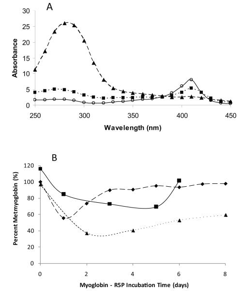
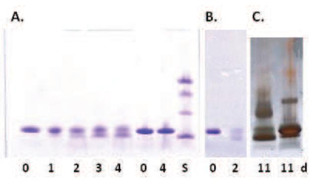
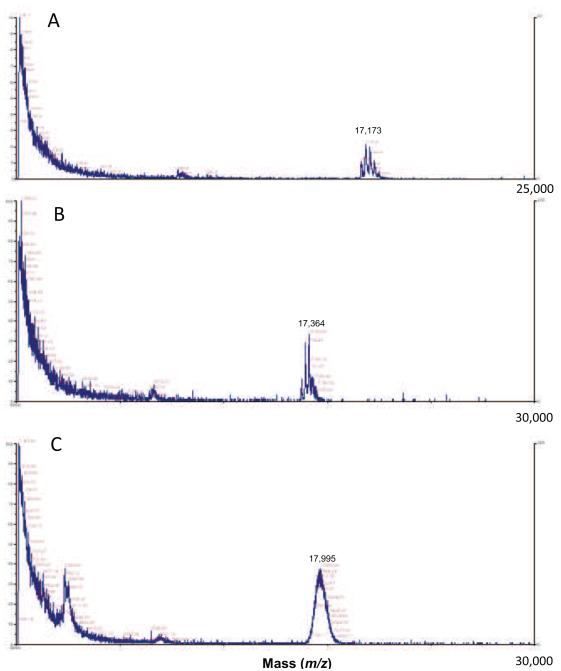
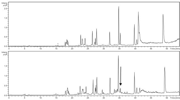
Glycation of horse heart metmyoglobin with d-ribose 5-phosphate (R5P), d-2-deoxyribose 5-phosphate (dR5P), and d-ribose with inorganic phosphate at 37°C generates an altered protein (Myo-X) with increased SDS-PAGE mobility. The novel protein product has been observed only for reactions with the protein myoglobin and it is not evident with other common sugars reacted over a 1 week period. Myo-X is first observed at 1-2 days at 37°C along with a second form that is consistent in mass with that of myoglobin attached to several sugars. MALDI mass spectrometry and other techniques show no evidence of the cleavage of a peptide from the myoglobin chain. Apomyoglobin in reaction with R5P also exhibited this protein form suggesting its occurrence was not heme-related. While significant amounts of O(2)(-) and H(2)O(2) are generated during the R5P glycation reaction, they do not appear to play roles in the formation of the new form. The modification is likely due to an internal cross-link formed during a glycation reaction involving the N-terminus and an internal amine group; most likely the neighboring Lys133. The study shows the unique nature of these common pentose sugars in spontaneous glycation reactions with proteins.
Copyright © 2011 Elsevier Inc. All rights reserved.
Figures
References
-
- Wautier JL, Schmidt AM. Protein glycation: a firm link to endothelial cell dysfunction. Circ. Res. 2004;95:233–238. - PubMed
-
- Cussimanio BL, Booth AA, Todd P, Hudson BG, Khalifah RG. Unusual susceptibility of heme proteins to damage by glucose during non-enzymatic glycation. Biophys. Chem. 2003;105:743–755. - PubMed
-
- Catalano CE, Choe YS, de Montellano P.R. Ortiz. Reactions of the protein radical in peroxide-treated myoglobin. Formation of a heme-protein cross-link. J. Biol. Chem. 1989;264:10534–10541. - PubMed
-
- Nagababu E, Rifkind JM. Reaction of hydrogen peroxide with ferrylhemoglobin: superoxide production and heme degradation. Biochemistry. 2000;39:12503–12511. - PubMed
-
- Roy A, Sen S, Chakraborti AS. In vitro nonenzymatic glycation enhances the role of myoglobin as a source of oxidative stress. Free Radic. Res. 2004;38:139–146. - PubMed
Publication types
MeSH terms
Substances
Grants and funding
LinkOut - more resources
Full Text Sources
