. 2011 Mar 4;144(5):675-88.
doi: 10.1016/j.cell.2011.01.031.

TLR signaling is required for Salmonella typhimurium virulence

Affiliations

TLR signaling is required for Salmonella typhimurium virulence

Nicholas Arpaia et al. Cell. .

Abstract

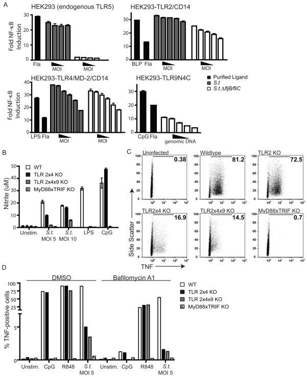
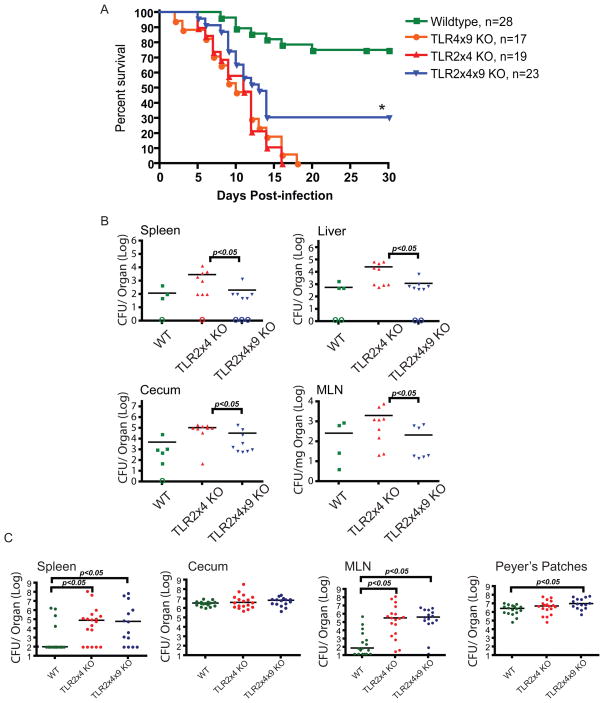
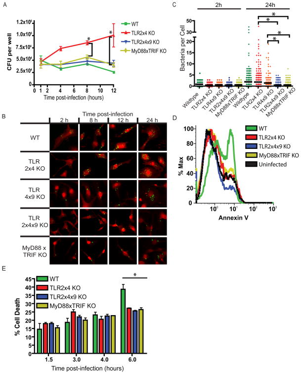
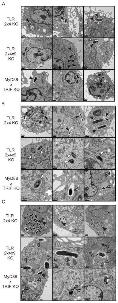
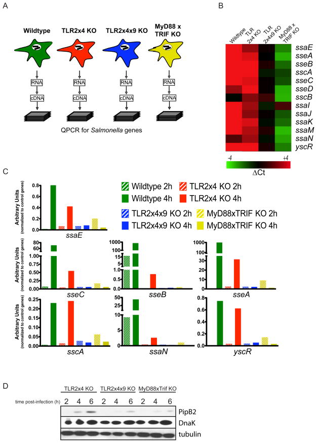
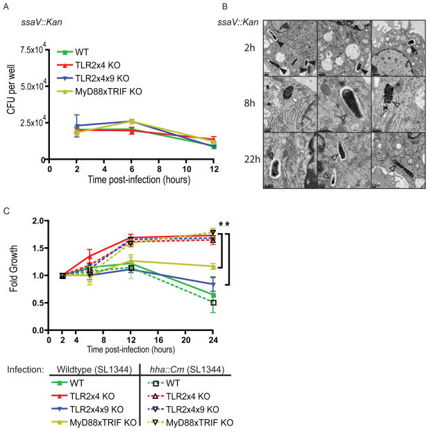
Toll-like receptors (TLRs) contribute to host resistance to microbial pathogens and can drive the evolution of virulence mechanisms. We have examined the relationship between host resistance and pathogen virulence using mice with a functional allele of the nramp-1 gene and lacking combinations of TLRs. Mice deficient in both TLR2 and TLR4 were highly susceptible to the intracellular bacterial pathogen Salmonella typhimurium, consistent with reduced innate immune function. However, mice lacking additional TLRs involved in S. typhimurium recognition were less susceptible to infection. In these TLR-deficient cells, bacteria failed to upregulate Salmonella pathogenicity island 2 (SPI-2) genes and did not form a replicative compartment. We demonstrate that TLR signaling enhances the rate of acidification of the Salmonella-containing phagosome, and inhibition of this acidification prevents SPI-2 induction. Our results indicate that S. typhimurium requires cues from the innate immune system to regulate virulence genes necessary for intracellular survival, growth, and systemic infection.


Figures

Figure 1
Figure 1. Multiple TLRs recognize products of S. typhimurium
(A) HEK293 cells expressing the indicated TLRs or TLR accessory proteins together with an NF-κB luciferase reporter were treated with heat-killed wildtype LT2 S. typhimurium (S.t.), flagellin-deficient LT2 (S.t.ΔfljB/fliC), or genomic DNA isolated from flagellin-deficient LT2. Luciferase activity was measured after 8h. Relative MOI range: 100 to 3.125, DNA concentration: 375 ng/mL to 12.5 ng/mL. Data are representative of 2 independent experiments and shown as mean +/− SEM. LPS, lipopolysaccharide; Fla, flagellin; BLP, bacterial lipopeptide. (B) BMMs differentiated from the indicated mice were treated overnight with 100 U/mL recombinant IFN-γ and infected the next morning with wildtype S. typhimurium (SL1344) at the indicated MOI. Nitrite production was measured 36h post-infection by Griess assay. Data are representative of 3 independent experiments and presented as mean +/− SD. (C) BMMs were infected as in (B) at an MOI of 5 for 8h, followed by intracellular cytokine staining for TNF. Percent TNF-positive cells are indicated in each panel. Data are representative of 3 independent experiments. (D) BMMs pre-treated for 2h with bafilomycinA1 or DMSO vehicle were infected with S. typhimurium (SL1344) at an MOI of 5 for 6h. Cells were processed and stained for intracellular cytokine staining as in (C).
Figure 2
Figure 2. TLR2x4x9-KO mice are less susceptible to S. typhimurium than TLR2x4-KO mice
(A) Survival plots of mice orally inoculated with 1.6 × 10⁸ CFU S. typhimurium (SL1344) are shown. * p<0.005 by log-rank curve comparison test. Data are representative of at least 2 independent experiments (B) Groups of 8–10 week-old mice were orally inoculated with 1 × 10⁹ CFU S. typhimurium (SL1344). Four days post-infection organs were harvested and homogenized for colony enumeration. Data are representative of at least 3 independent experiments. (C) Groups of mice of the indicated genotype were orally inoculated with 2 × 10⁸ Y. entericolitica and CFU were measured in the indicated organs 3 days post-infection. Data presented are the combined results from 2 independent experiments. For (B) and (C), bars represent mean CFU of all mice, with data significance determined by Mann-Whitney U test. Open circles indicate mice for which no colonies were detected. MLN, mesenteric lymph node. See also Figure S2.
Figure 3
Figure 3. S. typhimurium is unable to replicate in TLR2x4x9-KO and MyD88xTRIF-KO BMMs
(A) BMMs derived from mice of the indicated genotypes were infected with S. typhimurium (SL1344) at an MOI of 1, and intracellular CFU were measured by gentamicin protection assay at the indicated timepoints. Data are presented as the average of 3 independent experiments +/− SEM. * p<0.05 by student t test comparing TLR2x4-KO to MyD88xTRIF-KO and TLR2x4x9-KO. (B) BMMs infected as described in (A) were fixed and permeablized at the indicated times post-infection followed by staining with anti-Salmonella LPS antibody (green) and wheat germ agglutinin (red). (C) Intracellular bacteria per cell were counted in random fields at the 2 and 24h time-point from z-stacked images as shown in (B). Data are representative of 2 independent experiments, p value determined by student t test (* p<0.05). (D) BMMs of the indicated genotypes were infected with S. typhimurium (SL1344) at an MOI of 5. 8h post-infection, cells were harvested and stained with Annexin V. Data are representative of 2 independent experiments. (E) BMMs were infected at an MOI of 10 with S. typhimurium (SL1344), and release of lactate dehydrogenase (LDH) was measured in supernatants at the indicated time-points. Data are presented as mean +/− SD and are representative of at least 2 independent experiments; p value determined by student t test (* p<0.05) comparing wildtype to all other genotypes. See also Figure S3.
Figure 4
Figure 4. S. typhimurium fails to form an SCV in TLR2x4x9-KO and MyD88xTRIF-KO BMMs
TLR2x4-KO, TLR2x4x9-KO, or MyD88xTRIF-KO BMMs were infected at an MOI of 10 with S. typhimurium (SL1344), and cells were fixed and processed for electron microscopy at 2h (A), 8h (B), and 22h (C) post-infection. Bacteria in intact vacuoles are shown with filled black arrowheads, cytosolic bacteria with filled white arrowheads, and bacteria that are degraded or have fused with lytic compartments are indicated with open black arrowheads. Micron bars are in the lower left corner of each panel. Three representative images are shown from different sections and independent infections.
Figure 5
Figure 5. S. typhimurium fails to upregulate and secrete SPI-2 effectors in TLR2x4x9-KO and MyD88xTRIF-KO BMMs
(A) Schematic of quantitative expression analyses for S. typhimurium genes. Total RNA was isolated from infected BMMs of the indicated genotypes followed by processing for quantitative RT-PCR (see Experimental Procedures). (B) Heat map of normalized expression data for SPI-2 genes in bacteria within BMMs of the indicated genotypes. For each BMM genotype, data are shown relative to the 2h timepoint. The gene designation is to the right of each row. (C) Relative induction of individual SPI-2 genes in bacteria isolated from BMMs of the indicated genotypes. Data are normalized to the average expression values of a set of control genes. The data presented in (B) and (C) represent the mean of 2 independent experiments (D) BMMs of the indicated genotypes were infected with an S. typhimurium strain (12032) expressing an HA-tagged allele of pipB2 expressed from the endogenous pipB2 locus. The presence of PipB2 in BMM lysates was detected by immunoprecipitation and immunoblot with anti-HA antibodies. Controls for number of BMM (tubulin) and bacteria (DnaK) are also shown. Data are representative of 3 independent experiments.
Figure 6
Figure 6. TLR-signaling is necessary for SPI-2 induction and intracellular growth
(A) BMMs of the indicated genotypes were infected (MOI of 1) with SPI-2 mutant S. typhimurium (SL1344 ssaV::Kan). Intracellular CFU were measured by gentamicin protection assay. (B) TLR2x4-KO BMMs were infected as in (A) followed by fixation and processing for electron microscopy. Bacteria in intact vacuoles are indicated by filled black triangles, cytosolic bacteria by filled white triangles, and bacteria that are degraded or have fused with lytic compartments by open black triangles. Three representative images from different sections and from independent infections are shown. (C) BMMs of the indicated genotypes were infected (MOI of 1) with wildtype S. typhimurium or a strain with constitutive SPI-2 expression (SL1344 hha::Cm). Intracellular CFU were measured via gentamicin protection assay at the indicated timepoints post-infection. Data are presented as mean fold over the first time point (to control for minor inoculum differences between strains) +/− SEM and are representative of 3 independent experiments. * p<0.05 by student t test comparing TLR2x4-KO to MyD88xTRIF-KO and TLR2x4x9-KO at the indicated timepoint.
Figure 7
Figure 7. TLR-dependent phagosomal acidification is required for SPI-2 expression
(A) BMMs pre-treated with each of the indicated inhibitors, were infected with an S. typhimurium strain (12032) expressing an HA-tagged allele of pipB2. PipB2 levels in BMM lysates were detected by immunoprecipitation and immunoblot with anti-HA antibodies at 2, 4, 6 and 8 hours post-infection. Immunoblots for tubulin and DnaK serve as loading controls for BMMs and bacteria, respectively. Data are representative of 2 independent experiments for wildtype cells and 3 independent experiments for TLR2x4-KO BMMs. (B) BMMs were infected with FITC-labeled S. typhimurium (SL1344) and the fluorescence intensities of individual bacteria excited at 490 nm or 440 nm were measured over time by live cell imaging. The fluorescence intensity ratio (490/440) reflects the pH within the phagosome (see Experimental Procedures). The plots presented represent the mean pH calculated from at least 35 independent bacteria in multiple imaging fields. The right panel shows the percent of bacteria at or below pH 6 at 30 minutes post-infection. See Experimental Procedures and Figure S4.

Comment in

References

    1. Akira S, Takeda K, Kaisho T. Toll-like receptors: critical proteins linking innate and acquired immunity. Nat Immunol. 2001;2:675–680. - PubMed
    1. Ayres JS, Schneider DS. A signaling protease required for melanization in Drosophila affects resistance and tolerance of infections. PLoS Biol. 2008;6:2764–2773. - PMC - PubMed
    1. Bader MW, Sanowar S, Daley ME, Schneider AR, Cho U, Xu W, Klevit RE, Le Moual H, Miller SI. Recognition of antimicrobial peptides by a bacterial sensor kinase. Cell. 2005;122:461–472. - PubMed
    1. Bafica A, Scanga CA, Feng CG, Leifer C, Cheever A, Sher A. TLR9 regulates Th1 responses and cooperates with TLR2 in mediating optimal resistance to Mycobacterium tuberculosis. J Exp Med. 2005;202:1715–1724. - PMC - PubMed
    1. Bellamy R, Ruwende C, Corrah T, McAdam KP, Whittle HC, Hill AV. Variations in the NRAMP1 gene and susceptibility to tuberculosis in West Africans. N Engl J Med. 1998;338:640–644. - PubMed

Publication types