. 2011 Mar 4;144(5):810-23.
doi: 10.1016/j.cell.2011.02.018.

Astrocyte-neuron lactate transport is required for long-term memory formation

Affiliations

Astrocyte-neuron lactate transport is required for long-term memory formation

Akinobu Suzuki et al. Cell. .

Abstract

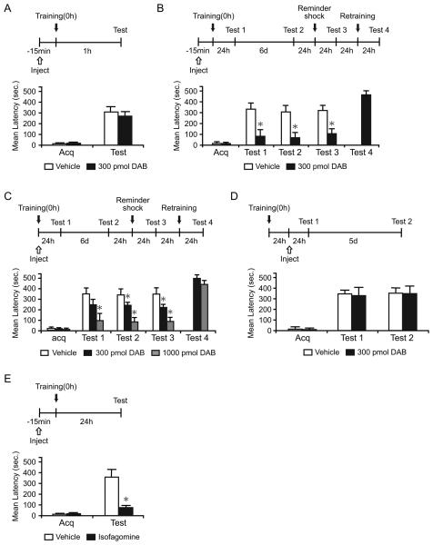
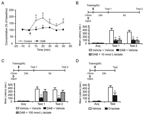
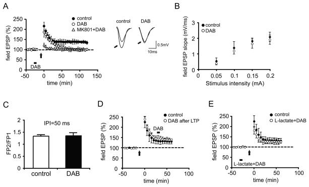
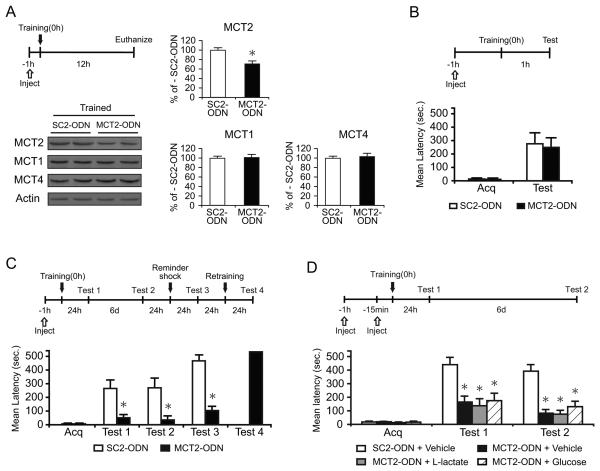
We report that, in the rat hippocampus, learning leads to a significant increase in extracellular lactate levels that derive from glycogen, an energy reserve selectively localized in astrocytes. Astrocytic glycogen breakdown and lactate release are essential for long-term but not short-term memory formation, and for the maintenance of long-term potentiation (LTP) of synaptic strength elicited in vivo. Disrupting the expression of the astrocytic lactate transporters monocarboxylate transporter 4 (MCT4) or MCT1 causes amnesia, which, like LTP impairment, is rescued by L-lactate but not equicaloric glucose. Disrupting the expression of the neuronal lactate transporter MCT2 also leads to amnesia that is unaffected by either L-lactate or glucose, suggesting that lactate import into neurons is necessary for long-term memory. Glycogenolysis and astrocytic lactate transporters are also critical for the induction of molecular changes required for memory formation, including the induction of phospho-CREB, Arc, and phospho-cofilin. We conclude that astrocyte-neuron lactate transport is required for long-term memory formation.


Figures

Figure 1
Figure 1. DAB and isofagomine disrupt long-term memory
Acquisition (Acq) and retention are expressed as mean latency ± SEM (in seconds, sec). Latency scores, n and detailed statistic are reported in Table S1. See also Figure S1. (A) Hippocampal injections of DAB 15 min before IA training had no effect on short-term memory tested at 1 hr after training (n=7/group). (B and C) Hippocampal injections of DAB 15 min before (B, n=11/group) or immediately after training (C, n=7-9/group) disrupted long-term memory at 24 hr (Test 1). The disruption persisted 7 days after training (Test 2), and memory did not recover after a reminder shock (Test 3). DAB-injected rats had normal retention after retraining (Test 4). (D) Hippocampal injections of DAB 24 hr after IA training did not affect long-term memory (n=8/group). (E) Hippocampal injections of isofagomine 15 min before training disrupted long-term memory (n=8-9/group). * p < 0.05.
Figure 2
Figure 2. L-lactate rescues the DAB-induced memory impairment
Lactate concentration, latency scores, n and detailed statistic are reported in Table S2. (A) Dorsal hippocampal extracellular lactate in freely moving rats infused with either vehicle or DAB. Baseline was collected for 20 min before training (0 min, ↑) and continued for 50 min. Training resulted in a significant increase in lactate levels compared to baseline (* p < 0.05) that was completely blocked by DAB (# p < 0.05). Data are expressed as % of baseline ± SEM (mean of the first 2 samples set 100%). See also Figure S2. (B-D) Acquisition (Acq) and retention are expressed as mean latency ± SEM (in seconds, sec). (B and C) Hippocampal injection of DAB or vehicle in combination with 10 nmol (B, n = 7/group), 100 nmol L-lactate (C, n = 12/group) or vehicle were performed 15 min before training and memory was tested at 24 hr. 100 nmol but not 10 nmol of L-lactate rescued the memory impairment by DAB (Test 1). The effect persisted at 7 days after training (Test 2). (D) Hippocampal injections of D-lactate 15 min before training disrupted long-term memory (n=7-8/group). * p < 0.05.
Figure 3
Figure 3. DAB impairs in vivo hippocampal LTP and the impairment is rescued by L-lactate
(A) Average Field EPSP data recorded for 120 min post-tetanus shows that DAB injection (bar) before high frequency stimulation (arrow) blocks LTP (p < 0.05 vs controls at 120 min, n = 4/group). LTP is abolished in animals receiving an intraperitoneal injection of the N-methyl-D-aspartate (NMDA) receptor antagonist MK-801 (3mg/kg) 30 min prior to tetanus (open triangles). Inset: Representative EPSP traces were recorded before and 120 min after (indicated by arrows) LTP induction. Left panel, control; right panel, in the presence of DAB. (B and C) No effect on the relationship between stimulus strength and the size of the postsynaptic response (input-output relationship, B, n = 4/group, p > 0.05) or paired-pulse facilitation (PPF, C, n = 4/group, p > 0.05). (D) DAB injected 30 min after high frequency stimulation did not affect synaptic potentiation (n = 4/group). (E) L-lactate reversed the blocking effect of DAB on LTP (n = 4/group). All data are expressed as mean values ± SEM.
Figure 4
Figure 4. MCT1 is induced by training. Disruption of MCT1 expression impairs long-term memory. Memory is rescued by L-lactate
Mean %, n, latency and detailed statistic are reported in Table S3. (A) MCTs expression performed on total (T) or synaptoneurosomal (S) extracts of dorsal hippocampus. Postsynaptic density-95 (PSD95) is used to show synaptoneurosmal enrichment. Glial fibrillary acidic protein (GFAP) is used as an astrocytic marker. (B) Examples and densitometric quantitative western blot analysis of MCT1, MCT2 and MCT4 carried out in dorsal hippocampal extracts from trained and untrained rats (n=6/group) injected with MCT1- or SC1-ODN and euthanized 12 hr after training. MCT1, but not MCT2 or MCT4, was significantly induced by training. This induction was selectively blocked by MCT1-ODN injection. Data are expressed as mean percentage ± SEM of the untrained-SC1-ODN (100%) mean values. All MCTs values were normalized to those of actin. (C) Examples and densitometric analysis of MCT1, MCT2 and MCT4 quantitative western blots of extracts from dorsal hippocampal punches taken around the needle placement from trained rats that received hippocampal MCT1- or SC1-ODN injections and were euthanized 12 h after training (n=4/group). Data are expressed as mean percentage ± SEM of trained-SC1-ODN (100%) mean values. All MCTs values were normalized to those of actin. (D-G) Memory acquisition (acq) and retention are expressed as mean latency ± SEM (in seconds, sec). (D) Hippocampal injections of MCT1-ODN 1 hr before training did not affect short-term memory (n=7/group). (E) Hippocampal injection of MCT1-ODN disrupted long-term memory. MCT1- or SC1-ODN were injected 1 hr before training and rats were tested 24 hr after training (Test 1) and 6 days later (Test 2). The memory disruption persisted at Test 2, and memory did not recover following a reminder shock (Test 3). MCT1-ODN amnesic rats showed normal retention after re-training (Test 4) (n=10-11/group). (F) L-lactate but not glucose rescued the memory impairment induced by blocking MCT1 expression (n=7-13/group). MCT1- or SC1-ODN were injected 1 hr before training. Llactate, glucose or vehicle (PBS) were injected 15 min before training. Rats were tested 24 hr after training (Test 1) and 6 days later (Test 2). (G) High concentration of glucose transiently rescued the MCT1-ODN-induced memory impairment (n = 8-11/group). MCT1- or SC1-ODN were injected 1 hr before training. 150 nmol of glucose or vehicle (PBS) were injected 15 min before training. Rats were tested 24 hr after training (Test 1) and 6 days later (Test 2). * p < 0.05.
Figure 5
Figure 5. Disruption of MCT4 expression also impairs long-term memory
Mean %, n, latency and detailed statistic are reported in Table S4. (A) Examples and densitometric analysis of MCT4, MCT1 and MCT2 quantitative western blots of extracts from dorsal hippocampal punches taken around the needle placement from trained rats that received hippocampal MCT4- or SC4-ODN injections and were euthanized 12 h after training (n=4/group). Data are expressed as mean percentage ± SEM of trained-SC4-ODN (100%) mean values. All MCTs values were normalized to those of actin. (B) Memory acquisition (acq) and retention are expressed as mean latency ± SEM (in seconds, sec). Hippocampal injection of MCT4-ODN disrupted long-term memory and L-lactate but not glucose, rescued it. MCT4- or SC4-ODN were injected 1 hr before training. L-lactate, glucose or vehicle (PBS) were injected 15 min before training. Rats were tested 24 hr after training (Test 1) and 6 days later (Test 2). (n=10-12/group). * p < 0.05
Figure 6
Figure 6. Disruption of MCT2 expression impairs long-term memory
Mean %, n, latency and detailed statistic are reported in Table S5. (A) Examples and densitometric analysis of MCT2, MCT1 and MCT4 quantitative western blots of extracts from dorsal hippocampal punches taken around the needle placement from trained rats that received hippocampal MCT2- or SC2-ODN injections and were euthanized 12 h after training (n=4/group). Data are expressed as mean percentage ± SEM of trained-SC2-ODN (100%) mean values. All MCTs values were normalized to those of actin. (B-D) Acquisition (acq) and retention are expressed as mean latency ± SEM (in seconds, sec). (B) Hippocampal injections of MCT2-ODN 1 hr before training did not affect short-term memory tested 1 hr later (n=8/group). (C) Hippocampal injections of MCT2-ODN disrupted long-term memory. MCT2- or SC2-ODN were injected 1 hr before training. Rats were tested 24 hr after training (Test 1) and 6 days later (Test 2). The memory disruption persisted at Test 2, and memory did not recover following a reminder foot shock (Test 3). The amnesic rats that received the MCT2-ODN showed normal retention after re-training (Test 4) (n = 8/group). (D) Neither L-lactate nor glucose rescued the memory impairment induced by MCT2 disruption (n = 6-8/group). MCT2- or SC2-ODN were injected 1 hr before training. Llactate, glucose or vehicle (PBS) were injected 15 min before training. Rats were tested 24 hr after training (Test 1) and 6 days later (Test 2). * p < 0.05.
Figure 7
Figure 7. Training-induced increase of Arc, pCREB and pcofilin are completely blocked by DAB and significantly rescued by L-lactate
Mean %, n and detailed statistic are reported in Table S6. (A-F) Examples (A) and densitometric western blot analysis of Arc (B), pCREB (C), CREB (D), pcofilin (E) and cofilin (F) performed on dorsal hippocampal extracts from trained and untrained rats injected 15 min before training with vehicle, DAB+vehicle or DAB+L-lactate and euthanized 30 min (for Arc) or 20 hr (for all other markers) after training. See also Figure S3. Arc (B), pCREB (C) and pcofilin (E) expression were significantly increased after training. This increase was completely blocked by DAB and rescued by L-lactate. There is no change in expression of CREB (D) and cofilin (F) across samples (n=4-7/group). Data are expressed as mean percentage ± SEM of untrained, vehicle-injected control (100%) mean values. All proteins values were normalized to those of actin. * p < 0.05.

Comment in

  • Astrocytes: powering memory.
    Bezzi P, Volterra A. Bezzi P, et al. Cell. 2011 Mar 4;144(5):644-5. doi: 10.1016/j.cell.2011.02.027. Cell. 2011. PMID: 21376229

References

    1. Alberini CM. Transcription factors in long-term memory and synaptic plasticity. Physiol. Rev. 2009;89:121–145. - PMC - PubMed
    1. Bamburg JR, McGough A, Ono S. Putting a new twist on actin: ADF/cofilins modulate actin dynamics. Trends Cell. Biol. 1999;9:364–370. - PubMed
    1. Bozdagi O, Nagy V, Kwei KT, Huntley GW. In vivo roles for matrix metalloproteinase-9 in mature hippocampal synaptic physiology and plasticity. J. Neurophysiol. 2007;98:334–344. - PMC - PubMed
    1. Bramham CR, Worley PF, Moore MJ, Guzowski JF. The immediate early gene arc/arg3.1: regulation, mechanisms, and function. J. Neurosci. 2008;28:11760–11767. - PMC - PubMed
    1. Brooks GA. Cell-cell and intracellular lactate shuttles. J. Physiol. 2009;587:5591–5600. - PMC - PubMed

Publication types