. 2011 Mar 11;88(3):306-16.
doi: 10.1016/j.ajhg.2011.02.001. Epub 2011 Mar 3.

Excess of de novo deleterious mutations in genes associated with glutamatergic systems in nonsyndromic intellectual disability

Collaborators, Affiliations

Excess of de novo deleterious mutations in genes associated with glutamatergic systems in nonsyndromic intellectual disability

Fadi F Hamdan et al. Am J Hum Genet. .

Erratum in

  • Am J Hum Genet. 2011 Apr 8;88(4):516

Abstract

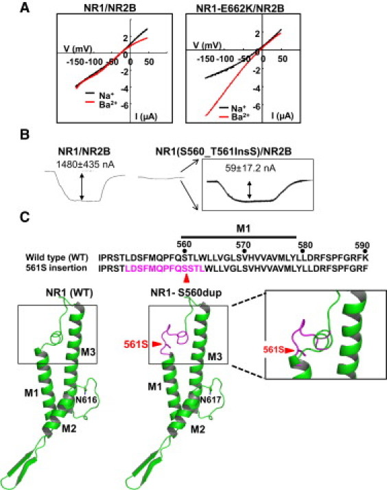
Little is known about the genetics of nonsyndromic intellectual disability (NSID). We hypothesized that de novo mutations (DNMs) in synaptic genes explain an important fraction of sporadic NSID cases. In order to investigate this possibility, we sequenced 197 genes encoding glutamate receptors and a large subset of their known interacting proteins in 95 sporadic cases of NSID. We found 11 DNMs, including ten potentially deleterious mutations (three nonsense, two splicing, one frameshift, four missense) and one neutral mutation (silent) in eight different genes. Calculation of point-substitution DNM rates per functional and neutral site showed significant excess of functional DNMs compared to neutral ones. De novo truncating and/or splicing mutations in SYNGAP1, STXBP1, and SHANK3 were found in six patients and are likely to be pathogenic. De novo missense mutations were found in KIF1A, GRIN1, CACNG2, and EPB41L1. Functional studies showed that all these missense mutations affect protein function in cell culture systems, suggesting that they may be pathogenic. Sequencing these four genes in 50 additional sporadic cases of NSID identified a second DNM in GRIN1 (c.1679_1681dup/p.Ser560dup). This mutation also affects protein function, consistent with structural predictions. None of these mutations or any other DNMs were identified in these genes in 285 healthy controls. This study highlights the importance of the glutamate receptor complexes in NSID and further supports the role of DNMs in this disorder.


Figures

Figure 1
Figure 1
Impact of p.Thr99Met on KIF1A Subcellular Localization EGFP-tagged motor domain (MD) constructs of KIF1A were expressed in cultured hippocampal neurons and visualized by immunofluorescence staining with an anti-EGFP. The MD of wild-type (WT) KIF1A (KIF1A-MD) accumulated in the axons (arrow heads), in contrast to KIF1A-MD-T99M or KIF1A-MD-T312M. The neuronal cell bodies and dendrites were stained with anti-MAP2 antibody.
Figure 2
Figure 2
Impact of GRIN1 p.Glu662Lys and p.Ser560dup on NMDAR Function Xenopus oocytes were coinjected with mRNAs of NR2B and either wild-type NR1 or mutated NR1 (E662K or S560dup). (A) Current-voltage curves of Na+ and Ba2+ currents. I-V curves for Glu+Gly-activated currents were measured by voltage ramps with Na saline and Ba saline. (B) Inward current induced by 10 μM glycine and 10 μM glutamate (Vh = −70 mV) in oocytes coinjected with NR2B and either wild-type NR1 or NR1-S560dup mRNAs. Inset: enlargement of the mutant NR1 signal. Values are means ± standard error of the mean (SEM) of eight independent experiments. (C) Conformational change of NR1 caused by p.Ser560dup. Top: amino acid sequences flanking the M1 transmembrane region are shown. The position of p.Ser560dup, resulting in the addition of a Ser561, is shown and indicated by an arrow head. Bottom: a 3D representation of the rat NR1 receptor subunit (WT and NR1-S560dup). M1, M2, and M3 represent transmembrane regions constituting the channel pore. The entrance to the channel pore (boxed) is significantly altered by the p.Ser560dup mutation.
Figure 3
Figure 3
Functional Impact of the p.Pro854Ser EPB41L1 Mutation (A) Indicated plasmid combinations (wild-type 4.1N [WT], 4.1N-P854S [P854S], myc-GluR) were cotransfected in HEK293 cells, and extracted proteins were immunoprecipitated with an anti-myc antibody and then analyzed by immunoblotting with anti-myc or anti-4.1N antibodies. The amount of coIP of 4.1N was reduced in the cells transfected with the 4.1N-P854S mutant. Right: the quantification of coIP experiment (mean ± SEM, n = 3, p < 0.05, t test). (B) Hippocampal neurons were cotransfected with GFP-GluR1 and the indicated 4.1N constructs. Surface GluR1 was stained by anti-GFP antibody in nonpermeabilized conditions and visualized by staining with a secondary anti-rabbit IgG (Alexa 568). Right: the quantification of GluR1 surface expression in presence of mutant or wild-type 4.1N (signal with WT adjusted to 1; mean ± SEM, n = 4, two-tailed Student's t test, p < 0.05)
Figure 4
Figure 4
Functional Impact of the p.Val143Leu CACNG2 Mutation (A) Indicated combinations of expression plasmids (wild-type myc-tagged stargazin [myc-Stg], myc-tagged stargazin-V143L [V143L], GFP-GluR1, GFP-GluR2) were cotransfected in HEK293 cells, and extracted proteins were immunoprecipitated with an anti-myc antibody and then analyzed by immunoblotting with anti-myc or anti-GFP antibodies. The amount of coIP of AMPARs (both GluR1 and GluR2) with Stg was reduced in the Stg-V143L transfected cells. Right: the quantification of the coIP experiments (mean ± SEM, n = 3, p < 0.05, t test). (B) Hippocampal neurons were transfected with GFP-GluR1 and indicated Stg constructs at DIV19 and stained with an anti-GFP (to detect surface GluR1) in nonpermeabilized conditions at DIV20. Stg-V143L results in reduced surface and total GluR1. Right: quantification of staining results (mean ± SEM, n = 4, ∗∗p < 0.01, p < 0.05, t test). (C) Hippocampal neurons were cotransfected with the indicated Stg constructs and GFP at DIV19, and miniature EPSCs were recorded at DIV22/23 from GFP-expressing neurons. Recordings were done in voltage-clamp mode at 65 mV in the presence of an extracellular solution containing 0.5 μM tetrodotoxin (TTX)/100 μM picrotoxin to detect excitatory mEPSC events. Expression of Stg-V143L mutant decreases both mEPSC amplitude and frequency. Right: results quantification (mean ± SEM, n = 4, p < 0.05, t test, ∗∗∗p < 0.001, t test).

References

    1. Kaufman L., Ayub M., Vincent J.B. The genetic basis of non-syndromic intellectual disability: A review. J. Neurodev. Disord. 2010;2:182–209. - PMC - PubMed
    1. Ropers H.H. Genetics of early onset cognitive impairment. Annu. Rev. Genomics Hum. Genet. 2010;11:161–187. - PubMed
    1. Laumonnier F., Cuthbert P.C., Grant S.G. The role of neuronal complexes in human X-linked brain diseases. Am. J. Hum. Genet. 2007;80:205–220. - PMC - PubMed
    1. Pocklington A.J., Cumiskey M., Armstrong J.D., Grant S.G. The proteomes of neurotransmitter receptor complexes form modular networks with distributed functionality underlying plasticity and behaviour. Mol. Syst. Biol. 2006;2:23. - PMC - PubMed
    1. Collins M.O., Husi H., Yu L., Brandon J.M., Anderson C.N., Blackstock W.P., Choudhary J.S., Grant S.G. Molecular characterization and comparison of the components and multiprotein complexes in the postsynaptic proteome. J. Neurochem. 2006;97(Suppl 1):16–23. - PubMed

Publication types

MeSH terms