. 2011 Jun;152(6):1263-1271.
doi: 10.1016/j.pain.2011.01.054. Epub 2011 Mar 4.

Leptin enhances NMDA-induced spinal excitation in rats: A functional link between adipocytokine and neuropathic pain

Affiliations

Leptin enhances NMDA-induced spinal excitation in rats: A functional link between adipocytokine and neuropathic pain

Yinghong Tian et al. Pain. 2011 Jun.

Abstract

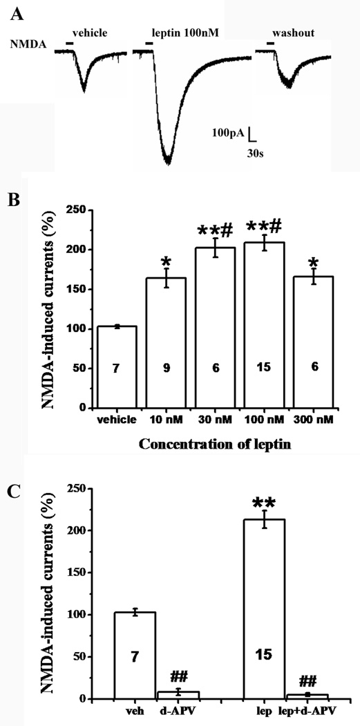
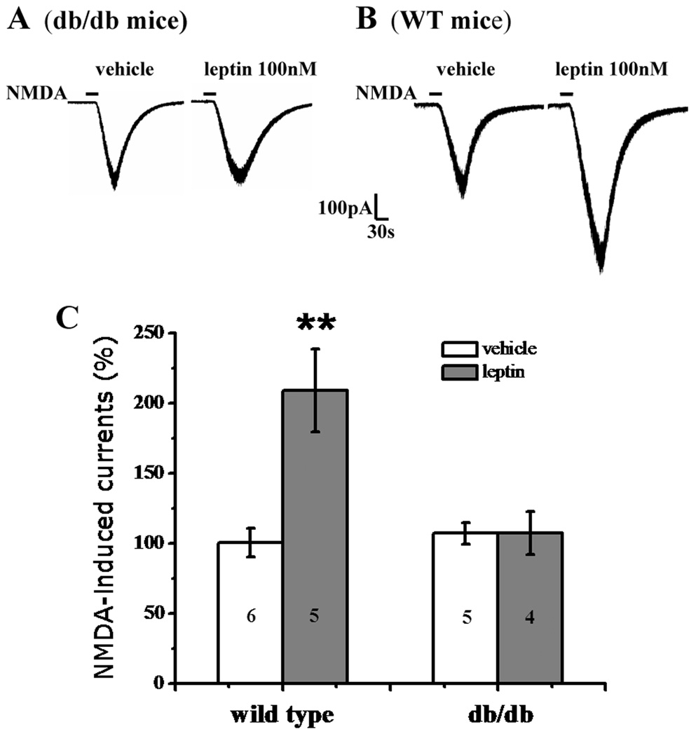
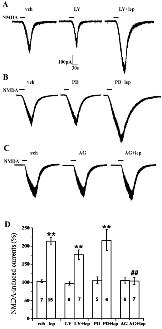
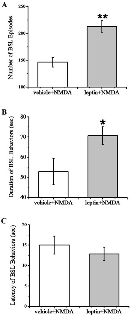
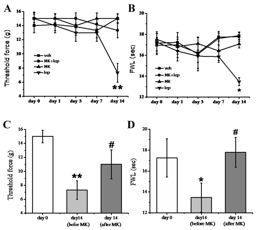
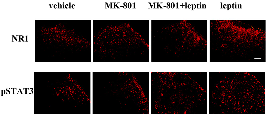
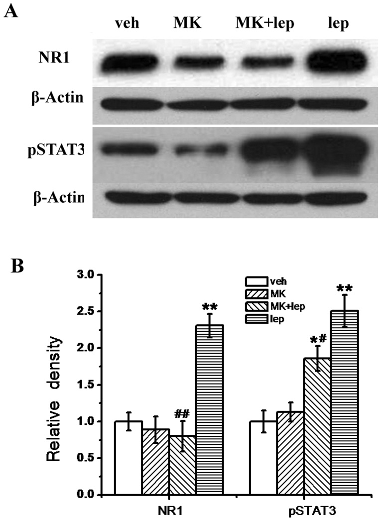
Recent studies have shown that leptin (an adipocytokine) played an important role in nociceptive behavior induced by nerve injury, but the cellular mechanism of this action remains unclear. Using the whole-cell patch-clamp recording from rat's spinal cord slices, we showed that superfusion of leptin onto spinal cord slices dose-dependently enhanced N-methyl-d-aspartate (NMDA) receptor-mediated currents in spinal cord lamina II neurons. At the cellular level, the effect of leptin on spinal NMDA-induced currents was mediated through the leptin receptor and the JAK2/STAT3 (but not PI3K or MAPK) pathway, as the leptin effect was abolished in leptin receptor-deficient (db/db) mice and inhibited by a JAK/STAT inhibitor. Moreover, we demonstrated in naïve rats that a single intrathecal administration of leptin enhanced spontaneous biting, scratching, and licking behavior induced by intrathecal NMDA and that repeated intrathecal administration of leptin elicited thermal hyperalgesia and mechanical allodynia, which was attenuated by the noncompetitive NMDA receptor antagonist MK-801. Intrathecal leptin also upregulated the expression of NMDA receptors and pSTAT3 within the rat's spinal cord dorsal horn, and intrathecal MK-801 attenuated this leptin effect as well. Our data demonstrate a relationship between leptin and NMDA receptor-mediated spinal neuronal excitation and its functional role in nociceptive behavior. Since leptin contributes to nociceptive behavior induced by nerve injury, the present findings suggest an important cellular link between the leptin's spinal effect and the NMDA receptor-mediated cellular mechanism of neuropathic pain. A functional link is demonstrated between leptin, an adipocytokine, and the cellular mechanisms of neuropathic pain via enhancement of function and expression of spinal N-methyl-d-aspartate receptors.


Conflict of interest statement

The authors declare no conflict of interest.

Figures

Fig. 1
Fig. 1. Enhancement by leptin of NMDA-induced currents
A, NMDA-induced current was enhanced in the presence of leptin (100 nM, 5min) and recovered at 30 minutes after leptin washout. B, The facilitatory effect of leptin on NMDA-induced currents was concentration-dependent. Leptin at 100 nM had the maximal enhancement effect. C, The enhancement by leptin of NMDA-currents was mediated through the NMDAR. The NMDAR antagonist d-APV (100µM) blocked NMDA-induced currents with or without leptin perfusion (n=6). Data are shown as mean±S.E. The number of cells in each group was shown inside each column. veh, vehicle; lep, leptin. *P < 0.05, **P < 0.01 versus vehicle; #P < 0.05 versus 10 nM and 300 nM leptin; $$P<0.01 versus before d-APV perfusion.
Fig. 2
Fig. 2. Lack of leptin effect in db/db mice
A, B, Representative traces showing the enhancement by leptin (100 nM, 5 min) of NMDA-induced currents in wild-type mice (B) but not in db/db mice (A). C, Histograms showing changes in NMDA currents in db/db mice and wild-type mice. Data are shown as mean±S.E. The number of cells in each group was shown inside each column. **P < 0.01 versus vehicle.
Fig. 3
Fig. 3. Role of the JAK2/STAT3 signal pathway in leptin-enhanced NMDA current
A, B, C, Representative traces showing the effect of the PI3K inhibitor LY-294,002 (A), MAPK inhibitor PD 98,059 (B), and JAK2/STAT3 inhibitor AG 490 (C) on the facilitation by leptin (100 nM, 5 min) of NMDA-induced currents. D, Histograms showing the effect of LY-294,002, PD 98,059, and AG 490 on the leptin effect on NMDA-induced currents. Data are shown as mean±S.E. The number of cells in each group was shown inside each column. veh, vehicle; lep, leptin; LY, LY-294,002; PD, PD 98,059; AG, AG 490. **P < 0.01 versus vehicle, ##P < 0.01 versus leptin.
Fig. 4
Fig. 4. Enhancement by leptin of NMDA-induced BSL behaviors
Intrathecal administration of NMDA (1 µg) elicited biting, scratching and licking (BSL) behaviors. The number of BSL episodes (A) and duration of BSL (B) induced by NMDA were increased by the pretreatment of 50 µg leptin (n=6, *P<0.05, ** P<0.01). (C) Pretreatment of leptin did not significantly change the latency of NMDA-induced BSL behaviors (n=6, P>0.05).
Fig. 5
Fig. 5. Effect of MK-801 on the role of leptin in hyperalgesia and allodynia
A, B: Intrathecal leptin (50 µg) in naïve rats, given once daily for 14 days, induced mechanical allodynia (A) and thermal hyperalgesia (B) when examined on day 14. The behavioral change was attenuated by coadministration of leptin with the NMDAR antagonist MK-801 (10 nmol) for 14 days. MK-801 (10 nmol) alone did not change the baseline nociceptive response. C, D: A single injection with MK-801 (10 nmol) on day 14 reversed both mechanical allodynia (C) and thermal hyperalgesia (D) induced by intrathecal leptin (50 µg, once daily) for 14 days. The data was obtained at 30 minutes after the MK-801 treatment. Data are shown as mean±S.E. veh, vehicle; lep, leptin; MK, MK-801. *P < 0.05, **P < 0.01 versus day 0; #P < 0.05 versus before the MK-801 injection on day 14. FWL: foot-withdrawal latency.
Fig. 6
Fig. 6. Leptin-induced spinal NR1 and pSTAT3 upregulation
Intrathecal leptin (50 µg) in naïve rats, given once daily for 14 days, upregulated the expression of the NR1 subunit and pSTAT3 within the spinal cord dorsal horn. The cellular changes were attenuated by coadministration of leptin with MK-801 (10 nmol) for 14 days. MK-801 alone did not change the baseline expression of NR1 and pSTAT3. For each panel, the upper right corner represents the dorsal part of the spinal cord. Scale bar: 100 µm.
Fig. 7
Fig. 7. Prevention by MK-801 of leptin-induced spinal NR1 and pSTAT3 upregulation
A, Western blot data showing the upregulation of NR1 and pSTAT3 in the spinal cord dorsal horn of rats receiving a 14-day intrathecal leptin (50 µg) treatment. B, Statistical data showing that MK-801 (10 nmol) prevented the upregulation of NR1 and pSTAT3 when coadministered with leptin for 14 days and MK-801 alone did not change the baseline expression of NR1 and pSTAT3. Data are shown as mean±S.E. *P < 0.05, P < 0.05, **P < 0.01 as compared with vehicle; #P < 0.05, ##P < 0.01 as compared with leptin. Veh, vehicle; lep, leptin; MK, MK-801; β-actin, loading control.

Similar articles

Cited by

References

    1. Ahima RS, Qi Y, Singhal NS, Jackson MB, Scherer PE. Brain adipocytokine action and metabolic regulation. Diabetes. 2006;55:S145–S154. - PubMed
    1. Banks WA. The many lives of leptin. Peptides. 2004;25:331–338. - PubMed
    1. Bjørbaek C, Kahn BB. Leptin signaling in the central nervous system and the periphery. Recent Prog Horm Res. 2004;59:305–331. - PubMed
    1. Bjorkman R, Hallman KM, Hedner J, Hedner T, Henning M. Acetaminophen blocks spinal hyperalgesia induced by NMDA and substance P. Pain. 1994;57:259–264. - PubMed
    1. Chen H, Charlat O, Tartaglia LA, Woolf EA, Weng X, Ellis SJ, Lakey ND, Culpepper J, Moore KJ, Breitbart RE, Duyk GM, Tepper RI, Morgenstern JP. Evidence that the diabetes gene encodes the leptin receptor: identification of a mutation in the leptin receptor gene in db/db mice. Cell. 1996;84:491–495. - PubMed

Publication types

MeSH terms