. 2011 Mar 22;21(6):439-48.
doi: 10.1016/j.cub.2011.01.073. Epub 2011 Mar 3.

Transport of germ plasm on astral microtubules directs germ cell development in Drosophila

Affiliations

Transport of germ plasm on astral microtubules directs germ cell development in Drosophila

Dorothy A Lerit et al. Curr Biol. .

Abstract

Background: In many organisms, germ cells are segregated from the soma through the inheritance of the specialized germ plasm, which contains mRNAs and proteins that specify germ cell fate and promote germline development. Whereas germ plasm assembly has been well characterized, mechanisms mediating germ plasm inheritance are poorly understood. In the Drosophila embryo, germ plasm is anchored to the posterior cortex, and nuclei that migrate into this region give rise to the germ cell progenitors, or pole cells. How the germ plasm interacts with these nuclei for pole cell induction and is selectively incorporated into the forming pole cells is not known.

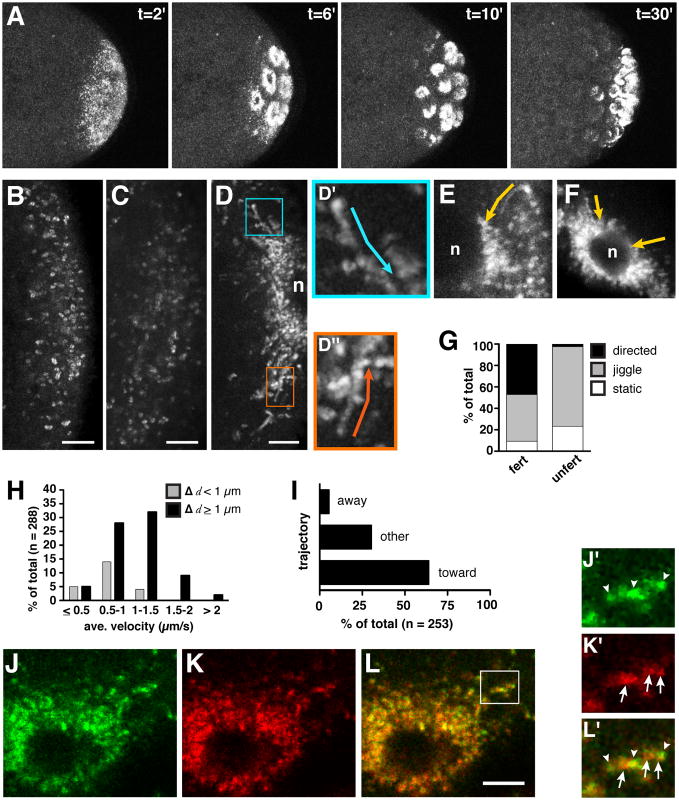
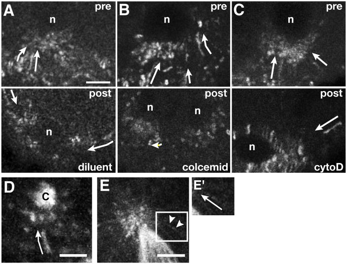
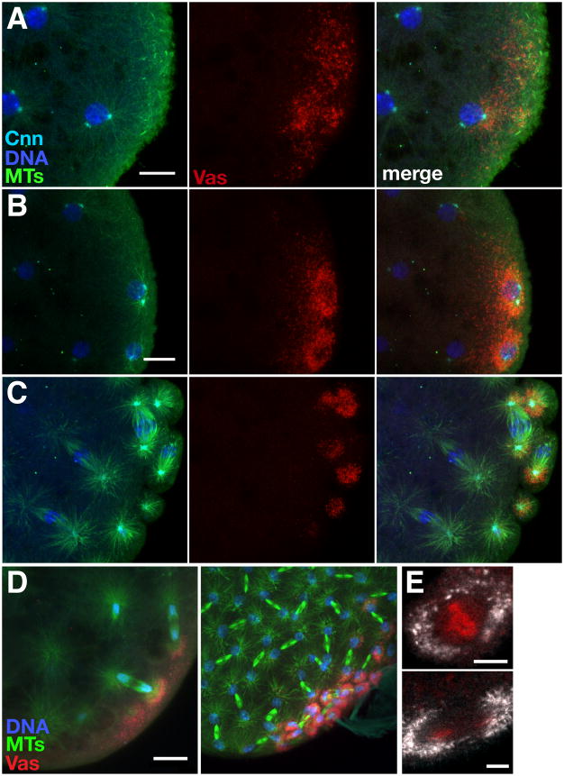
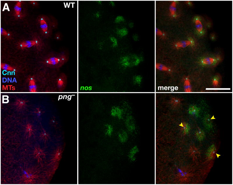
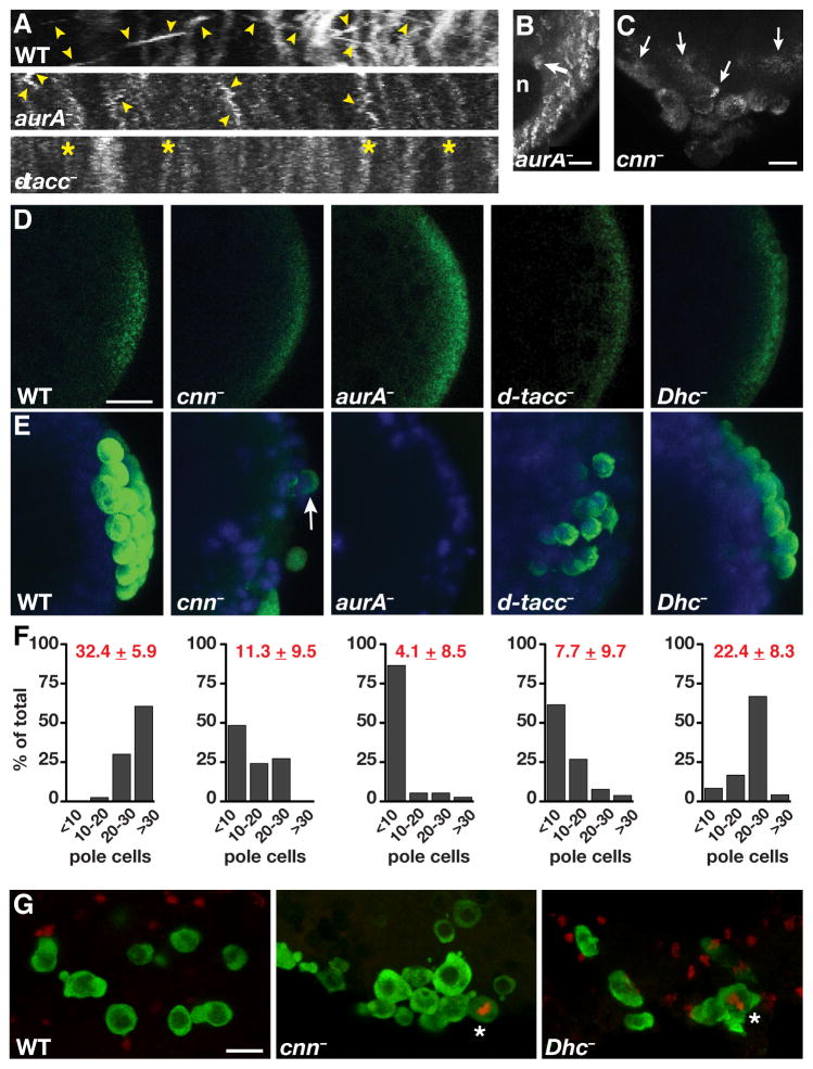
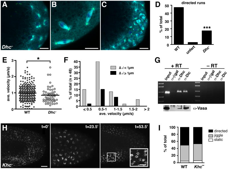
Results: Live imaging of two conserved germ plasm components, nanos mRNA and Vasa protein, revealed that germ plasm segregation is a dynamic process involving active transport of germ plasm RNA-protein complexes coordinated with nuclear migration. We show that centrosomes accompanying posterior nuclei induce release of germ plasm from the cortex and recruit these components by dynein-dependent transport on centrosome-nucleated microtubules. As nuclei divide, continued transport on astral microtubules partitions germ plasm to daughter nuclei, leading to its segregation into pole cells. Disruption of these transport events prevents incorporation of germ plasm into pole cells and impairs germ cell development.

Conclusions: Our results indicate that active transport of germ plasm is essential for its inheritance and ensures the production of a discrete population of germ cell progenitors endowed with requisite factors for germline development. Transport on astral microtubules may provide a general mechanism for the segregation of cell fate determinants.


Figures

Figure 1
Figure 1. Live imaging of nos mRNA and Vas protein during pole cell formation
(A) Two-photon excitation time-lapse imaging of the posterior region of an embryo expressing nos*GFP (anterior toward the left). Representative time points from Movie S1.1 are shown, where t=0 corresponds to the beginning of the time-series. (B–D) High resolution confocal time-lapse imaging of the posterior cortex of embryos expressing nos*GFP (anterior toward the left). 20–25 sequential frames spanning 6–8 seconds are superimposed so that moving particles appear as a trail of dots. (B) 0–1 hour old embryo (frames from Movie S1.2); (C) 1–2 hour old unfertilized embryo; (D) 1–2 hour old embryo with nucleus (n) at the posterior cortex (frames from Movie S1.3 Part 1); (D′ and D″) higher magnification of boxed regions in D showing sustained particle runs, with trajectories indicated by arrows. (E) Trail image showing nos*GFP detaching from the posterior cortex of a 1–2 hour embryo and traveling directly (arrow) toward a nucleus (n). Anterior is left. (F) Trail image of nos*GFP moving toward a nucleus at the anterior of a 1–2 hour old osk-bcd3′UTR embryo. (G) Percentage of particles undergoing movement in similarly aged 1–2 hour old fertilized embryos (fert; n=255 particles from 14 embryos) and unfertilized eggs (unfert; n=311 particles from 6 embryos). (H) Quantitation of average velocities and run lengths (Δ d) of motile particles (n=23 embryos). (I) Directionality of nos*GFP movement relative to nuclei labeled with H2AvD-mRFP (n=10 embryos) (see Movie S1.4). (J–L) Trail images from time-lapse analysis (Movie S1.5) of embryos expressing nos*GFP (J, green) and mCherry-Vas (K, red) show significant overlap (L, merge) as particles accumulate around a nucleus. Channels were imaged simultaneously under conditions where cross-talk is not detectable. (J′–L′) High power image of the region indicated by the box in (L). nos and Vas display heterogenous intensity profiles (arrowheads indicate predominant nos signal, arrows indicate predominant Vas signal). Scale bars: (B–D) 5 μm; (L) 2.5 μm. See also Figure S1 and Movies S1.1, S1.2, S1.3, S1.4, and S1.5.
Figure 2
Figure 2. Transport of nos*GFP is microtubule-dependent
Trail images from time-lapse movies of embryos with nos*GFP at the posterior cortex (anterior is up). (A–C) Pharmacological disruption experiments. Embryos were imaged immediately prior to (pre) and within 5 minutes after (post) injection of diluent (A), colcemid (B), or cytochalasin D (cytoD, C). Arrows indicate directed particle runs. (D) Trail image of nos*GFP (arrow) traveling toward a centrosome (c) labeled with GFP-Cnn. Six consecutive frames spanning two seconds of developmental time are superimposed. (E) Trail image of GFP-Vas moving on microtubules (arrowheads) labeled with GFP-α-tubulin. Time-lapse images spanning approximately 1.6 seconds are superimposed. The complete time sequence is shown in Movie S2.2. (E′) Magnification of the Vas particle in (E), with the trajectory indicated by the arrow. Scale bars: (A) 2.5 μm; (D) and (E) 5 μm. See also Movies S2.1 and S2.2.
Figure 3
Figure 3. Recruitment and segregation of germ plasm by centrosomal microtubules
(A–C) Confocal Z-series projections showing mCherry-Vas (Vas, red) together with microtubules (MTs, green), centrosomes (Cnn, cyan) and DNA (blue) at the posterior of embryos fixed prior to (A, B) and during (C) pole cell formation. Anterior is toward the left. Vas released from the cortex accumulates around astral microtubules as nuclei reach the posterior cortex (A) and remains associated with microtubules throughout nuclear divisions and pole cell formation (B, C). We have not yet been able to resolve whether the perinuclear ring appearance of the germ plasm (also Figure 1A) reflects the 3-dimensional organization of astral microtubules or whether there is a transient spreading of germ plasm around the nuclear periphery. (D) Anti-Vas (Vas, red) and anti-tubulin (MTs, green) immunofluorescence. Z-series projections spanning 20 μm, showing asymmetric accumulation of Vas around posterior nuclei (blue) with only one astral MT array in proximity to the posterior pole, restricting pole cell formation to the posterior. (E) Distribution of nos*GFP (white) relative to nuclei labeled with H2AvD-mRFP during metaphase (top) and anaphase (bottom). Scale bars: (A, B, D) 10 μm; (E) 5 μm.
Figure 4
Figure 4. Centrosomes are sufficient for release and recruitment of nos mRNA from the cortex
(A,B) Immunofluorescence detection of microtubules (MTs, red) and centrosomes (Cnn, cyan) in (A) wild-type (WT) or (B) png3318 (png) embryos expressing nos*GFP (green). Free centrosomes (arrowheads) and their microtubule arrays are sufficient to recruit nos. Scale bar: 20 μm. See also Figure S4.
Figure 5
Figure 5. Centrosomal microtubules mediate transport of germ plasm into pole cells for germ cell specification
(A) Representative kymographs from time-lapse images of 1–2 hour old mutant embryos expressing nos*GFP: aurA (aurA87Ac-3/aurA3); dtacc (dtacc1/Df(3R)110). Images were generated from a 300 × 200 pixel region of interest at the embryo posterior (ROI) and show particle movement over 35 frames, approximately 12 seconds of developmental time. The x axis spans 18.83 μm. Numerous linear runs (arrowheads) are detected throughout the imaging period in wild-type embryos. nos particle motility is severely reduced in aurA mutants, although some short localized runs are observed (arrowheads), and is eliminated in this d-tacc mutant embryo (asterisk). (B) Trail image of a 1–2 hour old aurA embryo showing reduced nos particle movement and minimal accumulation of nos around a nucleus (n; arrow). Twenty consecutive frames spanning 6.6 seconds are superimposed. (C) Z-series projection (30 μm) from a time-lapse sequence of a cnn (cnnHK21) embryo showing inefficient and irregular incorporation of nos*GFP into malformed pole cells (arrows). (D, E) Anti-Vas immunofluorescence (green) and DAPI-stained nuclei (blue) in 0–2 hour old (D) or 3–5 hour old (E) embryos. Note that a large number of unfertilized d-tacc embryos lack detectable localized Vas (data not shown). Vas is present at the posterior of mutant embryos prior to pole cell formation (D) but it often fails to associate with posterior nuclei and pole cells are often absent or severely reduced in number (E). Arrow indicates a pole cell with little germ plasm. (F) Quantification of pole cell number is shown below the corresponding genotypes in (E). The mean ± SD is indicated in red for each genotype: WT (n=40 embryos); cnn (n=33 embryos); aurA (n=37 embryos); d-tacc (n= 26 embryos); Dhc (Dhc6–10/Dhc6-6, n=24 embryos). (G) Confocal Z-series projections of migrating germ cells from stage 10–13 wild-type (WT), cnn, and Dhc (Dhc6–10/Dhc6-6) embryos, immunostained for Vas (green) and for the mitotic marker phospho-histone H3 Ser10 (red). Mitotically active germ cells (asterisks) were not detected in wild-type embryos (n = 37) but were found in cnn (n = 3/25) and Dhc (n=3/30) embryos. Similar results were obtained with d-tacc mutant embryos (n = 2/28) although these embryos were more difficult to stage due to developmental defects (data not shown). aurA mutant embryos could not be properly analyzed due to severe earlier developmental defects. Scale bars: (B) 2.5 μm; (C, D, G) 10 μm.
Figure 6
Figure 6. Germ plasm transport requires dynein, but not kinesin, during pole cell formation
(A–C) Immunofluorescence detection of microtubules (cyan), centrosomes (green), and DNA (blue) at the posterior of Dhc6–10/Dhc6-6 (Dhc) embryos. (A, B) 0–1 hour old embryos with centrosomes and astral microtubules visible during interphase (A) and mitosis (B). (C) 1–2 hour old embryo. (D) Quantitation of motile nos*GFP particles in 30 second time-lapse images from 1–2 hour old wild-type (WT) embryos (n = 255 particles from 14 embryos), similarly aged unfertilized eggs (n = 311 particles from 6 eggs), and 1–2 hour old Dhc6–10/Dhc6–12 (Dhc) embryos (n = 235 particles from 6 embryos). ***p < 0.001 as determined by the two-tailed Student’s t-test. (E) Scatter plot comparing the particle velocities in wild-type and Dhc6–10/Dhc6–12 embryos, with each data point representing a single motile particle. The solid line indicates the mean value (wild-type = 1.01 ± 0.44 μm/s; Dhc = 0.89 ± 0.48 μm/s). *p ≤ 0.05 as determined by the Wilcoxon rank-sum test. (F) Distribution of the average velocities and run lengths (Δd) of nos*GFP particles in Dhc6–10/Dhc6–12 mutant embryos. (G) Co-immunoprecipitation analysis. RT-PCR detects nos following immunoprecipitiation of embryo extracts with antibodies for dynein heavy chain (α-Dhc), dynein intermediate chain (α-Dic), or a control antibody to β-galactosidase (α-βgal). Reactions were performed with (+) or without (−) reverse transcriptase (RT). Specific enrichment of nos in anti-Dhc and anti-Dic immunoprecipitates was confirmed under nonsaturating RT-PCR conditions (Figure S6G). Immunoblotting for Vas is shown below the corresponding samples in (G). (H) Time course showing the distribution of GFP-Vas in the posterior half of a Khc27 germline clone embryo (Khc, anterior to the left). Before nuclear migration (t=0) Vas is dispersed over the entire cortex. Following nuclear migration and divisions (t=23.5′ and t=53.5′), Vas associates with nuclei. (I) Percentage of GFP-Vas particles undergoing movement in 1–2 hour old wild-type (WT) embryos (n = 189 particles from 4 embryos) and Khc embryos (n = 755 particles from 4 embryos). Scale bars: (A–C) 10 μm; (H) 20 μm, inset 10 μm. See also Figure S6 and Movies S6.1 and S6.2.

References

    1. Strome S, Lehmann R. Germ versus soma decisions: lessons from flies and worms. Science. 2007;316:392–393. - PubMed
    1. Mahowald AP. Assembly of the Drosophila germ plasm. Int Rev Cytol. 2001;203:187–213. - PubMed
    1. Foe VE, Alberts BM. Studies of nuclear and cytoplasmic behaviour during the five mitotic cycles that precede gastrulation in Drosophila embryogenesis. J Cell Sci. 1983;61:31–70. - PubMed
    1. Santos AC, Lehmann R. Germ cell specification and migration in Drosophila and beyond. Curr Biol. 2004;14:R578–589. - PubMed
    1. Gavis ER, Lehmann R. Localization of nanos RNA controls embryonic polarity. Cell. 1992;71:301–313. - PubMed

Publication types

MeSH terms