. 2011 Mar 22;21(6):473-9.
doi: 10.1016/j.cub.2011.02.003. Epub 2011 Mar 3.

Temporal control of contractile ring assembly by Plo1 regulation of myosin II recruitment by Mid1/anillin

Affiliations

Temporal control of contractile ring assembly by Plo1 regulation of myosin II recruitment by Mid1/anillin

Maria Almonacid et al. Curr Biol. .

Abstract

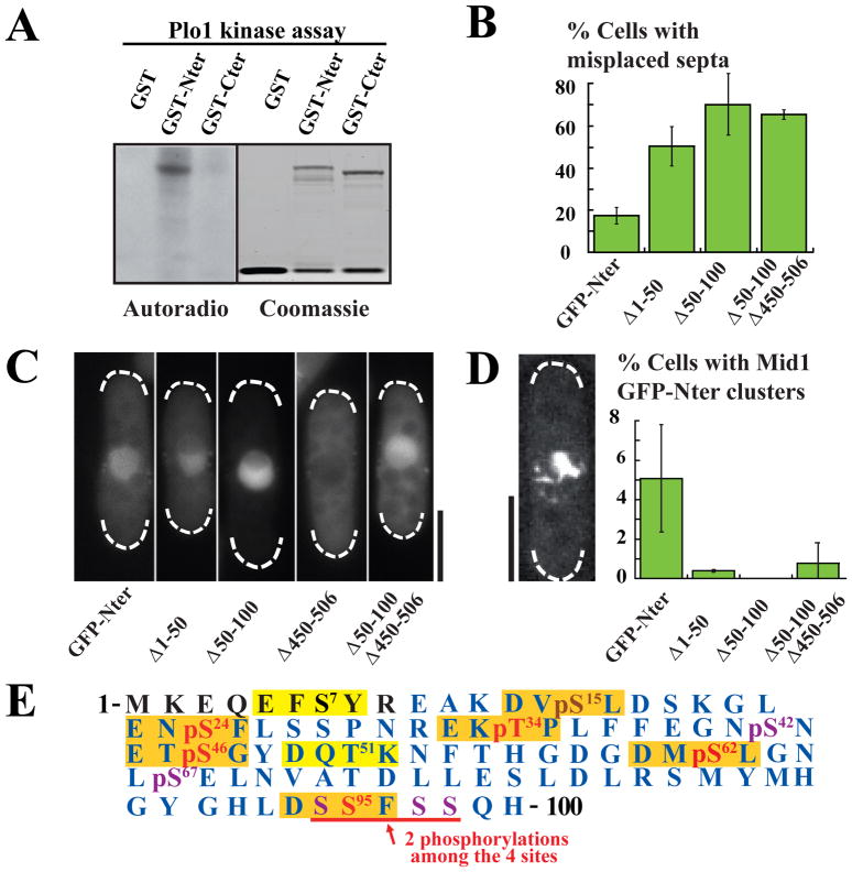
In eukaryotes, cytokinesis generally involves an actomyosin ring, the contraction of which promotes daughter cell segregation. Assembly of the contractile ring is tightly controlled in space and time. In the fission yeast, contractile ring components are first organized by the anillin-like protein Mid1 into medial cortical nodes. These nodes then coalesce laterally into a functional contractile ring. Although Mid1 is present at the medial cortex throughout G2, recruitment of contractile ring components to nodes starts only at mitotic onset, indicating that this event is cell-cycle regulated. Polo kinases are key temporal coordinators of mitosis and cytokinesis, and the Polo-like kinase Plo1 is known to activate Mid1 nuclear export at mitotic onset, coupling division plane specification to nuclear position. Here we provide evidence that Plo1 also triggers the recruitment of contractile ring components into medial cortical nodes. Plo1 binds at least two independent sites on Mid1, including a consensus site phosphorylated by Cdc2. Plo1 phosphorylates several residues within the first 100 amino acids of Mid1, which directly interact with the IQGAP Rng2, and influences the timing of myosin II recruitment. Plo1 thereby facilitates contractile ring assembly at mitotic onset.


Figures

Figure 1
Figure 1. Plo1 binding to Mid1 involves two sites and is favored by Cdc2 phosphorylation of threonine 517 within the RQST517PV consensus
A: Mid1 molecule (Nter, orange; Cter, blue). The consensus Plo1 binding site RQSTPV is detailed with phospho-T517 in red. B: In vitro binding of Plo1 PBD (MBP-Plo1-PBD) on Mid1 Nter and Cter (GST-Nter, aa 1–422; GST-Cter, aa 443–920). Top: MBP-Plo1-PBD and MBP (negative control) were revealed using α-MBP Abs. Bottom: loading controls (coomassie blue staining). C: Cdc2 kinase assay on Mid1 Cter. Left: Mid1 GST-Cter (aa 443–920) or GST-T517A-Cter were incubated with Cdc2 and Cdc2 kinase dead (kd). Phosphorylation is detected by 32P autoradiography. Right: loading controls (coomassie blue staining). D: Migration pattern of Mid1 Cter-GFP (aa 501–920) and Cter-T517A-GFP immunoprecipitated with an anti-GFP mAb and treated or not with CIP. WB: anti-GFP mAb. Red arrow: phospho-Cter-GFP. E: Coimmunoprecipitation of Mid1-12myc or Mid1-T517A-12myc with Plo1-GFP. IPs and WB for Plo1-GFP were performed with an anti-GFP mAb. Mid1 was detected using anti-Mid1 affinity purified Ab (α-Mid1). Negative controls: anti-GFP IPs on extracts from cells expressing untagged Plo1. Right: signal quantification. Raw signals of Mid1-12myc and Mid1-T517A-12myc in Plo1-GFP IP samples (left). Normalized signals relative to Mid1 and Mid1-T517A concentration in input (2.8 ratio; right). See also Figure S1.
Figure 2
Figure 2. Plo1 phosphorylates Mid1 Nter residues 1–100
A: Plo1 in vitro kinase assay on Mid1 GST-Nter (aa 1–422) or Mid1 GST-Cter (aa 443–920). Phosphorylation is detected by 32P autoradiography (left). Right: loading controls (coomassie blue staining). B: Percentage of misplaced septa in Mid1 GFP-Nter, GFP-Nter Δ1–50, Δ50–100 and Δ50–100Δ450–506 mutants. Cells were grown at 30°C. Error bars: SD, 9 independent counts of 100 cells. C: Localization of Mid1 GFP-Nter, GFP-Nter Δ1–50, Δ50–100, Δ450–506 and Δ50–100Δ450–506 deletion mutants expressed in mid1Δ cells during interphase. Bar: 5 μm. D: Percentage of GFP-Nter, GFP-NterΔ1–50, Δ50–100 and Δ50–100Δ450–506 cells assembling clusters of filaments upon Plo1 overexpression for 20 hours at 25°C. Error bars: SD, 3 independent experiments, n > 400. Left: GFP-Nter fluorescence in a cell showing a cluster. Bar: 5 μm. E: Phospho-map of Mid1 residues 1–100. Consensus Plo1 phosphorylation sites are boxed in orange when mutated in Mid1-6Ala mutant, or in yellow. Phosphosites detected by mass spectrometric analysis of mitotic GFP-Mid1 are shown in red in Plo1 consensus sites or purple. Sequence coverage is shown in blue. pS15 (in brown) was detected on Mid1 Nter phosphorylated in vitro by Plo1. See also Figure S2.
Figure 3
Figure 3. Mid1 activation by Plo1 controls the timing of Myosin II recruitment to medial cortical nodes
A: Time-lapse movies of cells deleted for endogenous mid1 expressing Mid1nsm-4GFP (top panel), Mid1nsm6Ala-4GFP (medium) or Mid1nsmΔ50–100-4GFP (bottom), Rlc1-mcherry and SPB component Sfi1-mRFP as a marker for mitotic entry. Time is in minutes and time 0 corresponds to SPB separation. Maximum projections of z stacks. Bars: 4 μm. B: Timing of Rlc1-mcherry cortical recruitment in same experiments as in A. Mid1nsm-4GFP: n=20. Mid1nsm6Ala-4GFP: n=39. Mid1nsmΔ50–100-4GFP: n=26. P value in Student t-test for equality between Mid1nsm6Ala or Mid1nsmΔ50–100 and Mid1nsm <10−10 and 10−12 respectively. C: Contractile ring assembly modes in same experiments as in A. Green: without filaments, blue: with filaments, red: with filaments starting at tips. Mid1nsm-4GFP: n=28. Mid1nsm6Ala-4GFP: n=62. Mid1nsmΔ50–100-4GFP: n=27. D: Percentage of misplaced septa in same strains as in A and in mid1Δ cells. Cells were grown at 30°C. Error bars: SD; 6 independent counts of 100 cells. See also Figure S3.
Figure 4
Figure 4. Mid1 residues 1–100 drive Mid1 localization at the contractile ring and interact with a Rng2 fragment
A: Localization of Cter-4GFP (left panel) and Mid1 1–100-Cter-4GFP (right panel) during mitosis in cells deleted for mid1 expressing Rlc1-mcherry and Sfi1-mRFP. Maximum projections of z stacks. Bar: 4 μm. B: Cell wall and septum staining (left) and percentage of normally placed septa (right) in same cells as in A, wild type (wt) or mid1Δ cells. Cells were grown at 30°C. Bar: 10 μm. Error bars: SD, 6 independent counts of 100 cells. C: In vitro binding of Rng2 C-termibrnal fragment (MBP-Rng2-1306-end) on Mid1 1–100 fragment (GST-Mid1 1–100). Proteins are stained with coomassie blue. GST and MBP are used as negative controls. Red star: MBP-Rng2-1306-end bound to GST-Mid1 1–100. D: Model for Mid1 activation by Plo1 at the onset of mitosis. In G2/M, Cdc2-dependent phosphorylation of Plo1 binding site RQST517PV favors Plo1 interaction with Mid1. Phosphorylation of Mid1 by Plo1 triggers Mid1 export from the nucleus, reinforces its localization at the medial cortex and couples the position of the division plane to nuclear position. Plo1 phosphorylation of domain 1–100, which interacts with a C-terminal fragment of Rng2, also triggers myosin II recruitment to medial cortical nodes and initiates the process of contractile ring assembly. See also Figure S4.

References

    1. Glotzer M. The molecular requirements for cytokinesis. Science. 2005;307:1735–1739. - PubMed
    1. Oliferenko S, Chew TG, Balasubramanian MK. Positioning cytokinesis. Genes Dev. 2009;23:660–674. - PubMed
    1. Pollard TD, Wu JQ. Understanding cytokinesis: lessons from fission yeast. Nat Rev Mol Cell Biol. 2010;11:149–155. - PMC - PubMed
    1. Roberts-Galbraith RH, Gould KL. Stepping into the ring: the SIN takes on contractile ring assembly. Genes Dev. 2008;22:3082–3088. - PubMed
    1. Chang F, Woollard A, Nurse P. Identification and characterization of fission yeast mutants defective in actin ring assembly and placement. J Cell Sci. 1996;109:131–142. - PubMed

Publication types

MeSH terms

LinkOut - more resources