. 2011 Mar 25;34(3):315-26.
doi: 10.1016/j.immuni.2011.01.013. Epub 2011 Mar 3.

A molecular basis for NKT cell recognition of CD1d-self-antigen

Affiliations

A molecular basis for NKT cell recognition of CD1d-self-antigen

Thierry Mallevaey et al. Immunity. .

Abstract

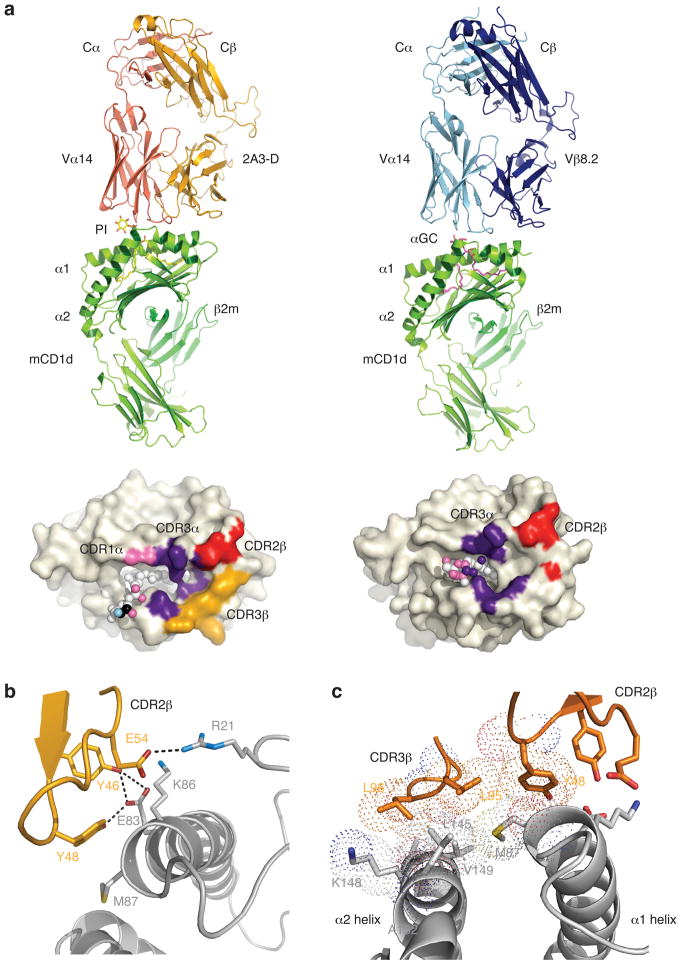
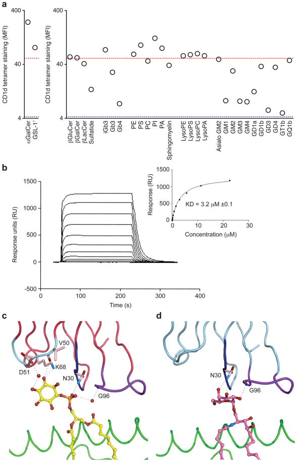
The antigen receptor for natural killer T cells (NKT TCR) binds CD1d-restricted microbial and self-lipid antigens, although the molecular basis of self-CD1d recognition is unclear. Here, we have characterized NKT TCR recognition of CD1d molecules loaded with natural self-antigens (Ags) and report the 2.3 Å resolution structure of an autoreactive NKT TCR-phosphatidylinositol-CD1d complex. NKT TCR recognition of self- and foreign antigens was underpinned by a similar mode of germline-encoded recognition of CD1d. However, NKT TCR autoreactivity is mediated by unique sequences within the non-germline-encoded CDR3β loop encoding for a hydrophobic motif that promotes self-association with CD1d. Accordingly, NKT cell autoreactivity may arise from the inherent affinity of the interaction between CD1d and the NKT TCR, resulting in the recognition of a broad range of CD1d-restricted self-antigens. This demonstrates that multiple self-antigens can be recognized in a similar manner by autoreactive NKT TCRs.


Figures

Figure 1
Figure 1
TCR CDR3β loop modulates αGC-CD1d tetramer binding. (a) Staining of hybridomas expressing the Vα14 TCRα chain paired with the Vβ8.2 chain in the context of the indicated CDR3β-Jβ rearrangement. The MFI of αGC-CD1d tetramer staining was determined for a narrow TCR gate. Data represent the mean + s.e.m. of two independent experiments. (b, c) Staining of hybridomas expressing the Vα14i TCRα chain paired with wild-type or CDR3β alanine substitutions of the DN32.D3 (b) or 24.8.A (c) TCRβ chains. The MFI of αGC-CD1d tetramer staining was determined for a narrow TCR gate and is normalized to wild-type MFI (set as 100%). Data represents the mean + s.e.m. of two independent experiments.
Figure 2
Figure 2
CDR3β loops and self-CD1d complex recognition. (a) Strategy used to identify CDR3β sequences that allow or not for self-CD1d complexes recognition. (b) Staining of hybridomas expressing the Vα14 TCRα chain paired with the indicated TCRβ chain. Data are representative of three independent experiments. (c) Enzyme-linked immunosorbent assay of interleukin-2 (IL-2) production by the indicated hybridomas in response to A20 cells transfected or not with mCD1d, in the presence of 10μg/ml 1B1 antibody or isotype control. Data represent the mean + s.e.m. of two independent experiments. (d) Staining by self-CD1d tetramers and inhibition of IL-2 production by 1B1 antibody for the hybridomas expressing self-CD1d-reactive NKT TCRs. Control, hybridoma expressing the Vα14i TCRα chain paired with the Vβ6 DO-11.10 TCRβ chain.
Figure 3
Figure 3
NKT TCR recognition of CD1d-PI. (a) Overview of the autoreactive 2A3-D NKT TCR-CD1d-PI complex (left hand side) compared to the Vβ8.2 NKT TCR-CD1d-αGC complex. Associated footprints on CD1d are shown underneath, and highlight the greater role of the CDR3β loop (gold) in the autoreactive complex. (b) Interactions mediated via the CDR2β loop in the 2A3-D NKT TCR-CD1d-PI complex (c) The two hydrophobic residues (Leu 95, Leu 96) in CDR3β act as a hydrophobic cap by shielding from solvent a surface exposed hydrophobic patch on CD1d. vdw radii shown as dots.
Figure 4
Figure 4
Mutational analysis of the 2A3-D NKT TCR. Staining of hybridomas expressing mutant versions (horizontal axes) of the Vα14i-2A3-Dβ TCRα (left) or TCRβ (right) with mouse CD1d tetramers loaded with naturally expressed self-antigens(s) (a) or αGC (b). Dark blue, CDR1α, magenta, CDR2α, green, CDR3α, light blue, CDR1β, light pink, CDR2β, yellow, CDR3β. WT, unsubstituted Vα14i-2A3-Dβ TCR (wild-type controls); control, Vα14i-Vβ6-DOβ TCR. ND, not done. The MFI of tetramer staining for each mutant was determined for a narrow TCR gate and is normalized to wild-type MFI (set as 100%). Data represent the mean normalized MFI + s.e.m. of at least two independent experiments.
Figure 5
Figure 5
Grafting of autoreactive CDR3β loop transfers autoreactivity. Staining of hybridomas expressing the Vα14 TCRα chain paired with the indicated Vβ chains in the context of the 2A3-D CDR3β. (a) Representative dot plots of three independent experiments. (b) The MFI of αGC-CD1d tetramer staining was determined for a narrow TCR gate. Data represent the mean + s.e.m. of three independent experiments. (c) Reactivity of the 2A3-D NKT TCR with CD1d molecules. Staining of the 2A3-D hybridoma with tetramers made of mouse CD1d monomers, or mouse CD1d:Ig complexes, produced in the indicated cell lines. Data are representative of three independent experiments.
Figure 6
Figure 6
Conserved recognition of self and exogenous antigens. Staining of hybridomas expressing the Vα14 TCRα chain paired with the indicated TCRβ chain. Data are representative of three independent experiments.
Figure 7
Figure 7
NKT TCR Recognition of CD1d-self Ag. (a) Antigen permissiveness by the NKT TCR. Staining of the hybridoma expressing the Vα14-Vβ8.2 TCR containing the CDR3β of 2A3-D with CD1d tetramers loaded with the indicated glycolipids or phospholipids (x axes). The MFI of CD1d tetramer staining was determined for a narrow TCR gate. Red dotted line, MFI with CD1d tetramer loaded with vehicle only (self-CD1d tetramer), blue dotted line, MFI with no addition of CD1d tetramer. Data represent the mean of two independent experiments. (b) Binding of the 2A3-D NKT TCR to mCD1d-PI as assessed by SPR. NKT TCR were injected over streptavidin immobilized mCD1d-PI and over an empty flow cell. Sensorgrams show the binding (response units, RU) of decreasing concentrations of TCR (22.5, 11.25, 5.625, 2.8125, 1.406 μM, 703, 351, 176, 87.9 43.9 nM) to the mCD1d-PI following subtraction of the control flow cell. Insets show saturation plots demonstrating equilibrium binding of NKT TCR to immobilized CD1d-PI. (c) Interactions between 2A3-D NKT TCR and PI (d) Interactions between Vβ8.2 NKT TCR and α-GC.

Comment in

  • The immutable recognition of CD1d.
    Adams EJ, López-Sagaseta J. Adams EJ, et al. Immunity. 2011 Mar 25;34(3):281-3. doi: 10.1016/j.immuni.2011.03.006. Immunity. 2011. PMID: 21435579

Similar articles

Cited by

References

    1. Arden B, Clark SP, Kabelitz D, Mak TW. Mouse T-cell receptor variable gene segment families. Immunogenetics. 1995;42:501–530. - PubMed
    1. Bellone M, Ceccon M, Grioni M, Jachetti E, Calcinoto A, Napolitano A, Freschi M, Casorati G, Dellabona P. iNKT cells control mouse spontaneous carcinoma independently of tumor-specific cytotoxic T cells. PLos One. 2010;5:e8646. - PMC - PubMed
    1. Bendelac A, Savage PB, Teyton L. The Biology of NKT Cells. Annu Rev Immunol. 2007;25:297–336. - PubMed
    1. Borg NA, Wun KS, Kjer-Nielsen L, Wilce MC, Pellicci DG, Koh R, Besra GS, Bharadwaj M, Godfrey DI, McCluskey J, Rossjohn J. CD1d-lipid-antigen recognition by the semi-invariant NKT T-cell receptor. Nature. 2007;448:44–49. - PubMed
    1. Brigl M, Brenner MB. How invariant natural killer T cells respond to infection by recognizing microbial or endogenous lipid antigens. Sem Immunol. 2009;22:79–86. - PubMed

Publication types