

Aldose reductase deficiency protects sugar-induced lens opacification in rats
- PMID: 21376710
- PMCID: PMC3103638
- DOI: 10.1016/j.cbi.2011.02.028
Aldose reductase deficiency protects sugar-induced lens opacification in rats
Abstract
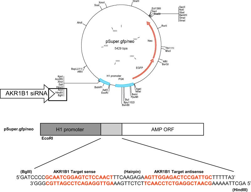
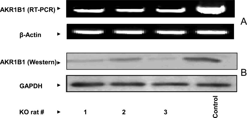
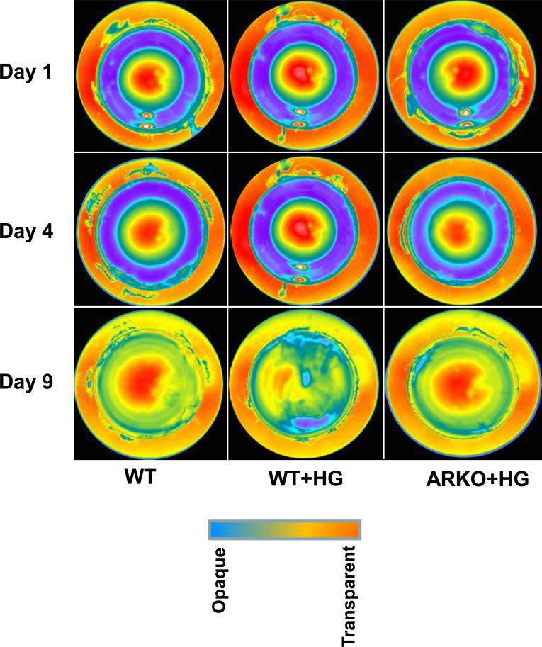
Aldose reductase (AKR1B1), which catalyzes the reduction of glucose to sorbitol and lipid aldehydes to lipid alcohols, has been shown to be involved in secondary diabetic complications including cataractogenesis. Rats have high levels of AKR1B1 in lenses and readily develop diabetic cataracts, whereas mice have very low levels of AKR1B1 in their lenses and are not susceptible to hyperglycemic cataracts. Studies with transgenic mice that over-express AKR1B1 indicate that it is the key protein for the development of diabetic complications including diabetic cataract. However, no such studies were performed in genetically altered AKR1B1 rats. Hence, we developed siRNA-based AKR1B1 knockdown rats (ARKO) using the AKR1B1-siRNA-pSuper vector construct. Genotyping analysis suggested that more than 90% of AKR1B1 was knocked down in the littermates. Interestingly, all the male animals were born dead and only 3 female rats survived. Furthermore, all 3 female animals were not able to give birth to F1 generation. Hence, we could not establish an AKR1B1 rat knockdown colony. However, we examined the effect of AKR1B1 knockdown on sugar-induced lens opacification in ex vivo. Our results indicate that rat lenses obtained from AKR1B1 knockdown rats were resistant to high glucose-induced lens opacification as compared to wild-type (WT) rat lenses. Biochemical analysis of lens homogenates showed that the AKR1B1 activity and sorbitol levels were significantly lower in sugar-treated AKR1B1 knockdown rat lenses as compared to WT rat lenses treated with 50mM glucose. Our results thus confirmed the significance of AKR1B1 in the mediation of sugar-induced lens opacification and indicate the use of AKR1B1 inhibitors in the prevention of cataractogenesis.
Copyright © 2011 Elsevier Ireland Ltd. All rights reserved.
Figures
References
-
- Hers HG. The mechanism of the transformation of glucose in fructose in the seminal vesicles. Biochim Biophys Acta. 1956;22:202–203. - PubMed
-
- Bhatnagar A, Srivastava SK. Biochemical aldose reductase: congenial and injurious profiles of an enigmatic enzyme. Med. Metab. Biol. 1992;48:91–121. - PubMed
-
- Dyck PJ, Thomas PK. Diabetic Neuropathy. W. B Saunders; Philadelphia: 1999.
-
- Brownlee M. Biochemistry and molecular biology of diabetic complications. Nature. 2001;414:813–820. - PubMed
-
- Beckman JA, Creager MA, Libby P. Diabetic and atherosclerosis: epidemiology, pathophysiology, and management. JAMA. 2002;287:2570–2581. - PubMed
Publication types
MeSH terms
Substances
Grants and funding
LinkOut - more resources
Full Text Sources
Medical
Molecular Biology Databases
Research Materials
