. 2011 Sep 1;714(1-2):1-10.
doi: 10.1016/j.mrfmmm.2011.02.006. Epub 2011 Mar 3.

Absence of polo-like kinase 3 in mice stabilizes Cdc25A after DNA damage but is not sufficient to produce tumors

Affiliations

Absence of polo-like kinase 3 in mice stabilizes Cdc25A after DNA damage but is not sufficient to produce tumors

David L Myer et al. Mutat Res. .

Abstract

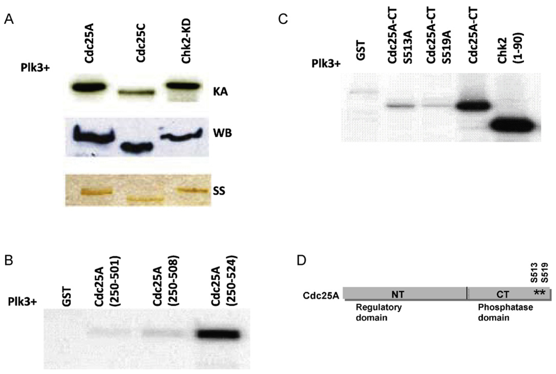
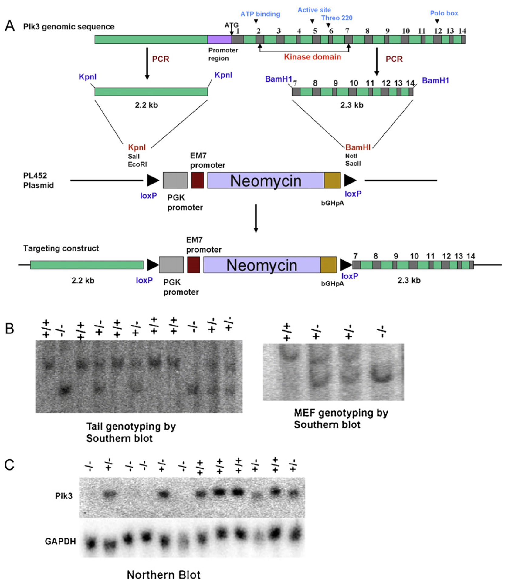
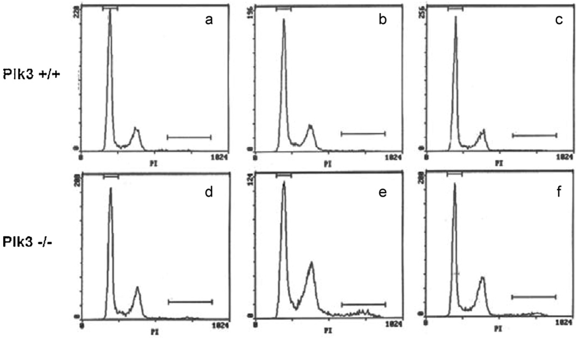
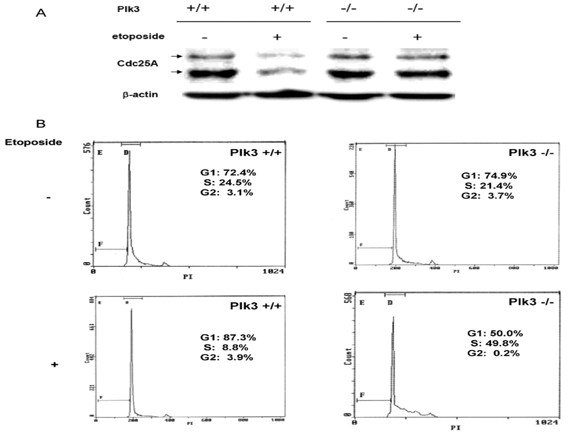
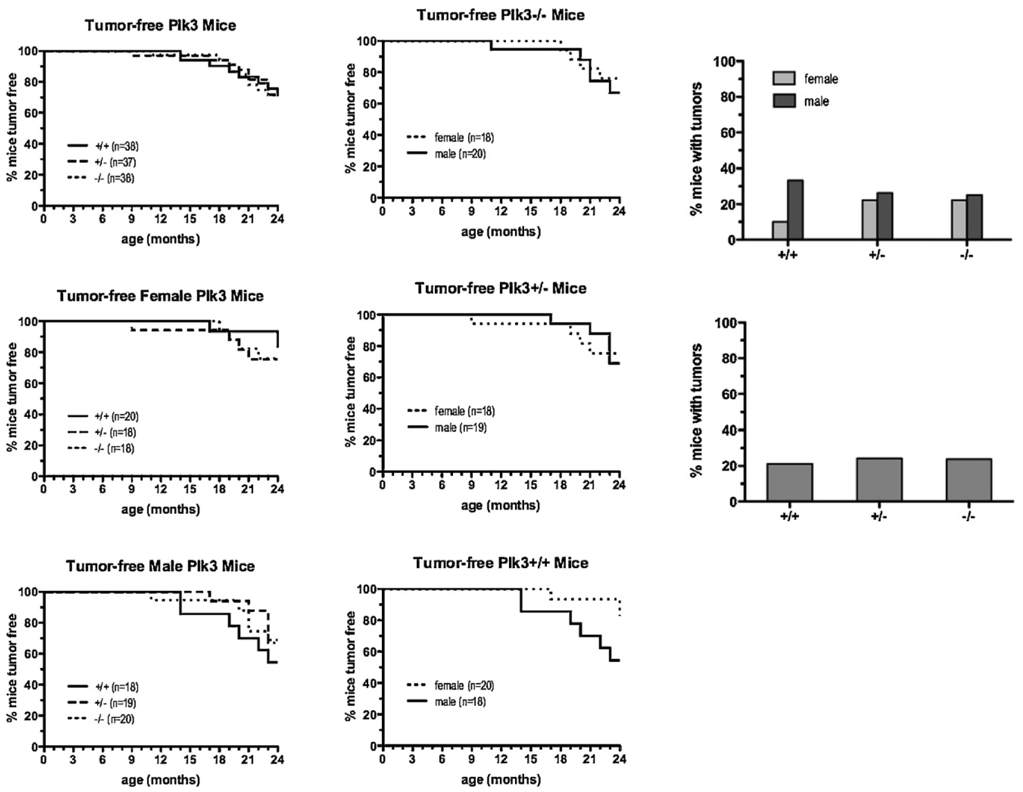
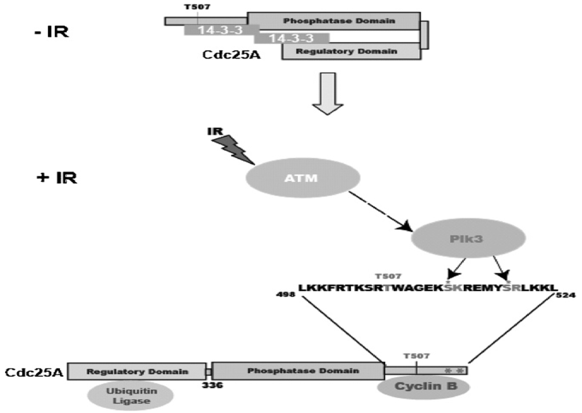
The polo-like kinases (Plks1-5) are emerging as an important class of proteins involved in many facets of cell cycle regulation and response to DNA damage and stress. Here we show that Plk3 phosphorylates the key cell cycle protein phosphatase Cdc25A on two serine residues in its cyclinB/cdk1 docking domain and regulates its stability in response to DNA damage. We generated a Plk3 knock-out mouse and show that Cdc25A protein from Plk3-deficient cells is less susceptible to DNA damage-mediated degradation than cells with functional Plk3. We also show that absence of Plk3 correlates with loss of the G1/S cell cycle checkpoint. However, neither this compromised DNA damage checkpoint nor reduced susceptibility to proteasome-mediated degradation after DNA damage translated into a significant increase in tumor incidence in the Plk3-deficient mice.


Conflict of interest statement

Conflict of interest

None.

Figures

Fig. 1.
Fig. 1.
Plk3 phosphorylates Cdc25A on serines 513 and 519 in vitro. (A) GST fusion Plk3 protein was expressed in bacteria and used to phosphorylate either Cdc25A or known substrates of Plk3 (Cdc25C and Chk2) in a kinase assay (KA). The amounts of used substrates were checked by both Western blot (WB) and silver staining (SS). (B) Deletion mutants of Cdc25A show that the phosphorylation sites are within the last 16 amino acids of Cdc25A protein. (C) Mutation of S513 or S519 on Cdc25A abolishes Plk3 phosphorylation activity. (D) A schematic representation of Cdc25A protein showing the location of the two phosphorylated sites on the C-terminal part of the protein.
Fig. 2.
Fig. 2.
Mutation of serines 513 or 519 to alanine in Cdc25A prevents proteasome-mediated degradation following IR treatment. HT1080 cells were transfected with pCDNA3 plasmid control, pcDNA3-myc-Cdc25A-WT, and with each of the pcDNA3-myc-Cdc25A mutants or left non-transfected (NT), and were subjected to IR treatment or left unchallenged. Cell lysates were probed with antibody to Myc. An incubation control where cells were taken out of the incubator while other cells were being treated with IR was also included. Beta-actin was used as the loading control.
Fig. 3.
Fig. 3.
Generation of a Plk3 knockout mouse. (A) A schematic representation of the targeting construct. (B) The targeted clones were identified by Southern blot (tail and MEF genotyping) and (C) Northern blot (mRNA expression).
Fig. 4.
Fig. 4.
Defect in cell cycle distribution in cells deficient in Plk3 following serum deprivation and release. Cells from wildtype mice and from mice lacking Plk3 were grown on regular medium (a and d) or deprived of serum and subsequently released for 24 h (b and e) or 36 h (c and f). Cell cycle distribution was analyzed by flow cytometry.
Fig. 5.
Fig. 5.
Thymocytes derived from Plk3−/− mice have a compromised G1/S checkpoint and are more resistant to degradation of Cdc25A than wildtype cells following DNA damage. Thymocytes derived from Plk3+/+ and Plk3−/− mice were left untreated or subjected to treatment with etoposide. (A) The Cdc25A level was analyzed by Western blot. (B) Cell cycle profile was determined by flow cytometry.
Fig. 6.
Fig. 6.
Development of spontaneous tumors in mice with wildtype and mutant Plk3. (A) Wildtype mice and mice heterozygous and homozygous for Plk3 were maintained for 24 months and monitored for palpable tumors twice a week. Mice were killed when tumors reached 1.0 cm in diameter or when mice appeared to be in distress or were found dead. In all cases, necropsy with histology of representative organs was performed, and tumors and tumor types were noted. (B) Comparison by gender of mice of each genotype that had developed spontaneous tumors by 24 months of age. (C) Comparison by gender (upper panel) or regardless of gender (lower panel) of mice of each genotype that had developed spontaneous tumors by 24 months of age.
Fig. 7.
Fig. 7.
A model depicting the regulation of Cdc25A degradation by Plk3. Plk3 phosphorylates serines 513 and 519 which are adjacent to the cyclinB/cdk1 docking site (K514 and R520). This phosphorylation, together with Chk1 phosphorylation of T507 allows 14–3–3 binding and prevents cyclinB/cdk1 from binding to Cdc25A. 14–3–3 binding leads to an open conformation of Cdc25A that allows binding of the ubiquitin ligase and subsequent degradation of the protein.

Similar articles

Cited by

References

    1. Glover DM, Hagan IM, Tavares AAM, Polo-like kinases: a team that plays throughout mitosis, Genes Dev. 12 (1998) 3777–3787. - PubMed
    1. Barr FA, Sillje HH, Nigg EA, Polo-like kinases and the orchestration of cell division, Nat. Rev. Mol. Cell Biol 5 (2004) 429–440. - PubMed
    1. Lane HA, Nigg EA, Antibody microinjection reveals an essential role for human polo-like kinase 1 (Plk1) in the functional maturation of mitotic centrosomes, J. Cell Biol 135 (1996) 1701–1713. - PMC - PubMed
    1. Qian YW, Erikson E, Li C, Maller JL, Activated polo-like kinase Plx1 is required at multiple points during mitosis in Xenopus laevis, Mol. Cell. Biol 18 (1998) 4262–4271. - PMC - PubMed
    1. Moutinho-Santos T, Sampaio P, Amorim I, Costa M, Sunkel CE, In vivo localisation of the mitotic POLO kinase shows a highly dynamic association with the mitotic apparatus during early embryogenesis in Drosophila, Biol. Cell 91 (1999) 585–596. - PubMed

Publication types

Substances