Review
. 2011 Jun 1;56(3):1437-52.
doi: 10.1016/j.neuroimage.2011.02.073. Epub 2011 Mar 3.

Functional connectivity MRI in infants: exploration of the functional organization of the developing brain

Affiliations
Review

Functional connectivity MRI in infants: exploration of the functional organization of the developing brain

Christopher D Smyser et al. Neuroimage. .

Abstract

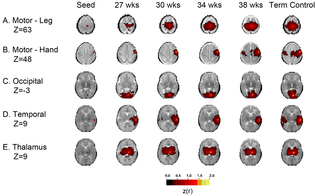
Advanced neuroimaging techniques have been increasingly applied to the study of preterm and term infants in an effort to further define the functional cerebral architecture of the developing brain. Despite improved understanding of the complex relationship between structure and function obtained through these investigations, significant questions remain regarding the nature, location, and timing of the maturational changes which occur during early development. Functional connectivity magnetic resonance imaging (fcMRI) utilizes spontaneous, low frequency (< 0.1 Hz), coherent fluctuations in blood oxygen level dependent (BOLD) signal to identify networks of functional cerebral connections. Due to the intrinsic characteristics of its image acquisition and analysis, fcMRI offers a novel neuroimaging approach well suited to investigation of infants. Recently, this methodology has been successfully applied to examine neonatal populations, defining normative patterns of large-scale neural network development in the maturing brain. The resting-state networks (RSNs) identified in these studies reflect the evolving cerebral structural architecture, presumably driven by varied genetic and environmental influences. Principal features of these investigations and their role in characterization of the tenets of neural network development during this critical developmental period are highlighted in this review. Despite these successes, optimal methods for fcMRI data acquisition and analysis for this population have not yet been defined. Further, appropriate schemes for interpretation and translation of fcMRI results remain unknown, a matter of increasing importance as functional neuroimaging findings are progressively applied in the clinical arena. Notwithstanding these concerns, fcMRI provides insight into the earliest forms of cerebral connectivity and therefore holds great promise for future neurodevelopmental investigations.


Conflict of interest statement

The authors declare that they have no competing financial interests or conflicts of interest.

Figures

Figure 1
Figure 1. Histological changes in the neocortex during prenatal development
The developing neocortex is composed of a series of embryonic cellular zones, with appearance (and in some cases subsequent resolution) dependent upon gestational age. Neurons are generated in the ventricular zone (VZ). The preplate (PP) is formed by migration of post-mitotic neurons from this region. Accumulation of intermediate progenitors creates the subventricular zone (SVZ). The cortical plate (CP) forms via ensuing neurogenesis and migration in an inside-out sequence, with cortical layers 2–6 emerging through subsequent rounds of cell division. The neurons divide the preplate into the marginal zone (MZ) (below the pial surface) and the subplate zone (SP) via radial migration. The marginal zone becomes layer 1 of mature cortex. The intermediate zone (IZ) lies between the proliferating layers and post-migratory neurons, providing the foundation for subsequent white matter development (Bystron, Blakemore et al. 2008). Images are schematic representations of the developing neocortex at approximately (A) embryonic day (E) 30, (B) E31–32, (C) E45, (D) E55, (E) gestational week 14, and (F) gestational week 18. Adapted with permission from Annual Reviews from (Kanold and Luhmann 2010).
Figure 2
Figure 2. Consistent fcMRI results in infants using varied acquisition parameters
Selection of image acquisition parameters can significantly affect fcMRI results, and limited investigation of the effect of these differences in this population has been performed. Lin et al used differing repetition time (TR) settings to acquire fcMRI data in subjects 0–2 years of age. Comparable results were identified for each group, suggesting minimal effects for TR time. (A, B) fcMRI correlation maps generated using seeds located in the right (R) and left (L) sensorimotor cortices for infants age two weeks and one year from data obtained using TR time of (A) 2 seconds and (B) 0.75 seconds. Note similar patterns of interhemispheric correlation between homotopic counterparts for each group. Z-score values are indicated by color bar on right (threshold value = 1.0). Adapted with permission from Williams and Wilkins Co. from (Lin, Zhu et al. 2008).
Figure 3
Figure 3. Gestational age specific atlases enable improved image registration
Spatial normalization to a common stereotactic space allows statistical comparison of inter- and intra-individual functional neuroimaging results. This procedure can be problematic in infants due to the rapid, regional-specific changes in cerebral anatomy which occur during early development. Use of atlas targets specific to narrow periods of development limits the impact of these effects, improving image registration and normalization. (A) Post menstrual age (PMA) specific targets generated for designated gestational age categories (27, 30, 34, and 38 weeks PMA and term-born infants; adult target provided for comparison) using a previously described iterative algorithm (Buckner, Head et al. 2004). Note progressive change in cerebral volume and gyration and sulcation patterns with advancing age. (B) Results transformed to common (adult) stereotactic space through application of age-specific, logarithmically averaged stretch. Adapted with permission from Oxford University Press from (Smyser, Inder et al. 2010).
Figure 4
Figure 4. Detection of intermittent head motion allowing exclusion of affected volumes with increased correlation strength
Subject motion generates artifact that cannot be corrected post-acquisition. This is of significant concern in investigations of infants who are prone to movement. However, head motion tends to occur sporadically in sleeping subjects and manifests as intermittent change (usually loss) of signal intensity. (A) Linear and rotational head movement during acquisition for a typical subject. Changes in mean signal intensity (most likely due to intermittent head motion) are reflected in the black trace, which reflects the reciprocal of the imaged mean whole brain intensity. (B) Results generated by head motion correction algorithm for same fMRI data. Frames exceeding an empirically determined rms tolerance (solid red line) are ignored in the correlation analysis. (C, D) fcMRI correlation maps generated using right thalamic seed for same subject without (C) and with (D) inclusion of results obtained from motion correction algorithm. Note increased interhemispheric connection strength with correction for subject movement. (C) and (D) show Fisher z-transformed correlation coefficients (color threshold value = 0.5). Adapted with permission from Oxford University Press from (Smyser, Inder et al. 2010).
Figure 5
Figure 5. Longitudinal neural network development in preterm infants
Average fcMRI correlation maps corresponding to varied seed locations. The images are organized in columns corresponding to PMA at time of imaging. The illustrated quantity is the group mean Fisher z-transformed correlation coefficient (color threshold value = 0.3) overlaid on the gestational age specific atlas. Each row shows the axial slice at the level of the seed region. Note age dependent network maturation. Included are representative networks identified utilizing seeds located in the (A) motor – leg (Z=63), (B) motor – hand (Z=48), (C) occipital (Z=−3), and (D) temporal (Z=9) cortices and (E) thalamus (Z=9). The right side of the image corresponds to the right side of the brain. Adapted with permission from Oxford University Press from (Smyser, Inder et al. 2010).

References

    1. Achard S, Bullmore E. Efficiency and cost of economical brain functional networks. PLoS Comput Biol. 2007;3(2):e17. - PMC - PubMed
    1. Achard S, Salvador R, et al. A resilient, low-frequency, small-world human brain functional network with highly connected association cortical hubs. J Neurosci. 2006;26(1):63–72. - PMC - PubMed
    1. Allendoerfer KL, Shatz CJ. The subplate, a transient neocortical structure: its role in the development of connections between thalamus and cortex. Annu Rev Neurosci. 1994;17:185–218. - PubMed
    1. Alstott J, Breakspear M, et al. Modeling the impact of lesions in the human brain. PLoS Comput Biol. 2009;5(6):e1000408. - PMC - PubMed
    1. Altaye M, Holland SK, et al. Infant brain probability templates for MRI segmentation and normalization. Neuroimage. 2008;43(4):721–730. - PMC - PubMed

Publication types

LinkOut - more resources