. 2011 May 15;353(2):186-93.
doi: 10.1016/j.ydbio.2011.02.025. Epub 2011 Mar 4.

Ascl3 knockout and cell ablation models reveal complexity of salivary gland maintenance and regeneration

Affiliations

Ascl3 knockout and cell ablation models reveal complexity of salivary gland maintenance and regeneration

Szilvia Arany et al. Dev Biol. .

Abstract

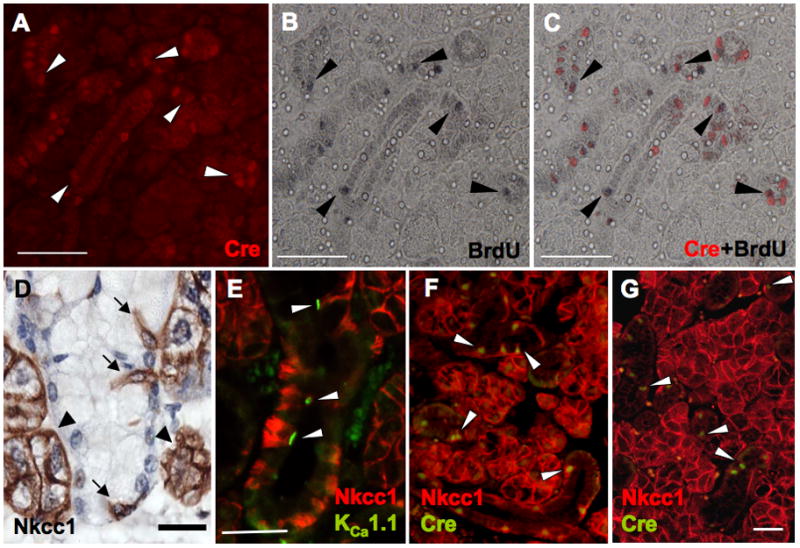
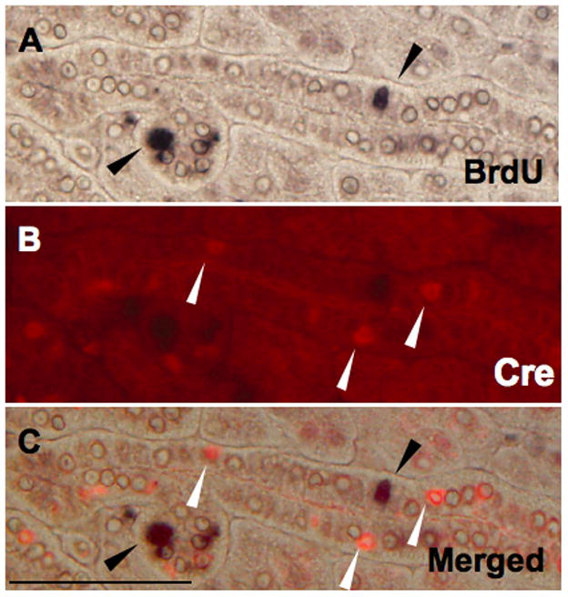
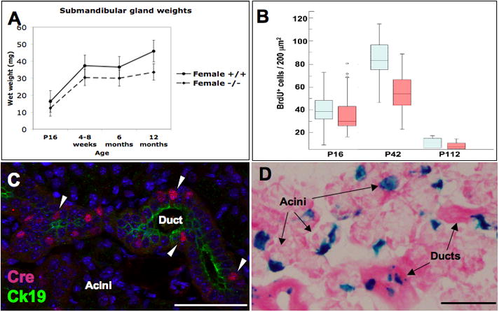
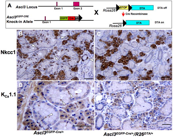
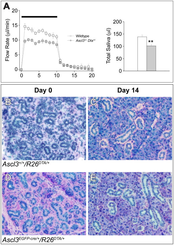
Expression of the transcription factor, Ascl3, marks a population of adult progenitor cells, which can give rise to both acinar and duct cell types in the murine salivary glands. Using a previously reported Ascl3(EGFP-Cre/+) knock-in strain, we demonstrate that Ascl3-expressing cells represent a molecularly distinct, and proliferating population of progenitor cells located in salivary gland ducts. To investigate both the role of the Ascl3 transcription factor, and the role of the cells in which it is expressed, we generated knockout and cell-specific ablation models. Ascl3 knockout mice develop smaller salivary glands than wild type littermates, but secrete saliva normally. They display a lower level of cell proliferation, consistent with their smaller size. In the absence of Ascl3, the cells maintain their progenitor function and continue to generate both acinar and duct cells. To directly test the role of the progenitor cells, themselves, in salivary gland development and regeneration, we used Cre-activated expression of diphtheria toxin (DTA) in the Ascl3-expressing (Ascl3+) cell population, resulting in specific cell ablation of Ascl3+ cells. In the absence of the Ascl3+ progenitor cells, the mice developed morphologically normal, albeit smaller, salivary glands able to secrete saliva. Furthermore, in a ductal ligation model of salivary gland injury, the glands of these mice were able to regenerate acinar cells. Our results indicate that Ascl3+ cells are active proliferating progenitors, but they are not the only precursors for salivary gland development or regeneration. We conclude that maintenance of tissue homeostasis in the salivary gland must involve more than one progenitor cell population.


Figures

Fig. 1
Fig. 1
Molecular characterization of Ascl3+ progenitor cells. Cell proliferation was detected through BrdU labeling in salivary glands from Ascl3EGFP-Cre/+ mice at P16 following a 2-hour chase period. Sections of submandibular glands were double-immunostained with antibodies to Cre recombinase and BrdU. A. Cre immunostaining identifies Ascl3+ cells in the ducts (arrowheads). B. BrdU immunostaining marks proliferating cells (arrowheads). C. Merged image shows that 50% of the BrdU-positive cells co-localize with those expressing Ascl3 (arrowheads). Scale bars = 100 μm. D. Antibody to the sodium-potassium-chloride transporter, Nkcc1, is localized to the basolateral membrane of all acinar cells (arrowheads) and to a subset of basally located duct cells (arrows) in the submandibular gland from a wild type male. Scale bar = 20 μm E. Double immunohistochemistry with antibodies to Nkcc1 (red) and KCa1.1 (green) reveals co-localization of the two proteins in a subset of the duct cells (arrowheads). Scale bar = 20 μm F. Double immunohistochemistry on sections from an Ascl3EGFP-Cre/+ heterozygote, using antibodies to Cre recombinase (green) and Nkcc1 (red) shows that the two proteins are co-localized in the same subset of duct cells, indicating that Ascl3+ cells co-express the Nkcc1 cotransporter. G. The same double immunohistochemistry with antibodies to Cre and Nkcc1, on sections from Ascl3 EGFP-Cre/EGFP-Cre knockout glands, shows that Nkcc1 expression in the Ascl3+ progenitor cells is significantly down regulated in the absence of Ascl3 transcription factor (arrowheads). In contrast, Nkcc1 expression in the acinar cells is unaffected. Scale bar = 50 μm
Fig. 2
Fig. 2
Ascl3-expressing cells are not label-retaining stem cells. Heterozygous Ascl3EGFP-Cre/+ pups were injected daily from P16 to P23 with BrdU. After an 8-week chase period, the salivary glands were removed, processed and analyzed by double immunohistochemistry, using antibodies for BrdU (A; black) and Cre recombinase (B; red). The sections, shown separately and as a merged image (C), include BrdU-positive, label-retaining cells (black arrowheads) and Ascl3-positive cells (white arrowheads) in the ducts. No co-localization of the two cell populations was observed. Scale bars = 50 μm
Fig. 3
Fig. 3
Salivary glands of Ascl3 EGFP-Cre/EGFP-Cre knockout mice. A. Submandibular glands of Ascl3 EGFP-Cre/EGFP-Cre mice are smaller than those of wild type littermates, from P16 throughout adulthood. Wet weights of submandibular glands isolated from Ascl3 +/+ and Ascl3 EGFP-Cre/EGFP-Cre knockout females are plotted for each age examined. Bars show standard error. (n≥6 for both genotypes) B. Cell proliferation is reduced in the submandibular glands of Ascl3 EGFP-Cre/EGFP-Cre knockout mice compared to Ascl3EGFP-Cre/+ heterozygotes. Box plot displays the data collected from counts done on 6 × 200 μm2 areas of sections from 4 animals of each genotype, following a 2-hour pulse of BrdU. Ascl3EGFP-Cre/+ heterozygotes (blue boxes); Ascl3 EGFP-Cre/EGFP-Cre knockout animals (red boxes). Boxes span from the first to the third quartiles of the count data. Median bars are indicated within the boxes. [P16: median Ascl3EGFP-Cre/+ = 38.5; median Ascl3 EGFP-Cre/EGFP-Cre = 30.0; P42: median Ascl3EGFP-Cre/+ = 82.5; median Ascl3 EGFP-Cre/EGFP-Cre = 54.0; P<0.01. P112: median Ascl3EGFP-Cre/+ = 10.0; median Ascl3 EGFP-Cre/EGFP-Cre= 7.0; P<0.05]. C. The differentiation status of the duct cells in Ascl3 EGFP-Cre/EGFP-Cre knockout glands was investigated using antibodies to Cre (red; arrowheads) and cytokeratin 19 (green), a luminal membrane marker of differentiated duct cells. Duct cell differentiation appears normal in the Ascl3 EGFP-Cre/EGFP-Cre knockout submandibular glands. All cell nuclei are stained with Topro-3 (blue). Confocal image. D. Lineage tracing of the progenitor cells in Ascl3 EGFP-Cre/EGFP-Cre knockout glands. LacZ staining demonstrates that, in the absence of the Ascl3 transcription factor, the progenitor cells remain bipotent and generate both duct and acinar cell progeny. Scale bars = 100 μm
Fig. 4
Fig. 4
Cell ablation of Ascl3+ progenitor cells. A. Ascl3EGFP-Cre/+/R26DTA/+ mice carry one copy of the EGFP-Cre cassette, and one copy of the silenced DTA gene inserted at the Rosa26 locus. Cre-mediated activation of DTA expression takes place only in Ascl3-expressing cells. DTA expression results in specific, cell-autonomous killing of the Ascl3-expressing cells. B. In control Ascl3EGFP-Cre/+ submandibular glands, Nkcc1 antibody is localized on the membranes of all acinar cells (brown) and on the subpopulation of Ascl3+ progenitor cells in the ducts (arrowheads). C. In submandibular glands of Ascl3EGFP-Cre/+/R26DTA/+ mice, all Nkcc1-expressing Ascl3+ progenitor duct cells are ablated, while Nkcc1 expression in the acinar cells (brown) is unaffected. D. In Ascl3EGFP-Cre/+ submandibular glands, antibody to KCa1.1 is localized at the apical surface of a subset of duct cells, shown earlier to co-localize with Nkcc1 and Ascl3 (arrowheads). E. In glands of Ascl3EGFP-Cre/+/R26DTA/+ mice, all cells expressing KCa1.1 are ablated, confirming the efficiency of the cell ablation model. Scale bars = 50 μm
Fig. 5
Fig. 5
Salivary gland function and regeneration in the cell ablation model. A. Salivary secretion in Ascl3EGFP-Cre/+/R26DTA/+ mice was measured by ex vivo submandibular gland perfusion after stimulation with 0.3 μm carbachol and 5 μm isoproterenol. In comparison to wild type mice, both the flow rate (μL/min) (left) and level of total secretion (μL in 20 minutes) (right) were significantly reduced in the glands of Ascl3EGFP-Cre/+/R26DTA/+ mice. However, when corrected for the differences in total submandibular gland weight, the secretory rates of Ascl3EGFP-Cre/+/R26DTA/+ mice are nearly equivalent to those of wild type mice. (n=8 Ascl3+/+/R26+/+; n=9 Ascl3EGFP-Cre/+/R26DTA/+) *P=0.0010 (t-test) B–E. Periodic acid-Schiff/Alcian blue staining of sections from submandibular glands after ductal ligation, followed by recovery for the time indicated. B. Section of Ascl3+/+/R26DTA/+ control mouse submandibular gland, taken at day 0 of recovery, shows the almost complete loss of acinar cells following 10 days of ductal ligation. Ducts predominate within the gland. C. Section of control Ascl3EGFP-Cre/+/R26+/+ mouse submandibular gland, taken at day 14 of recovery, shows repopulation of the acinar compartment between the ducts. D. Section of Ascl3EGFP-Cre/+/R26DTA/+ submandibular gland, taken at day 0 of recovery, also shows the loss of acinar cells following 10 days of ductal ligation. Spaces between the ducts appear empty or contain fibrotic tissue. E. Section of Ascl3EGFP-Cre/+/R26DTA/+ submandibular gland, lacking Ascl3+ progenitor cells, taken at day 14 of recovery, shows that repopulation of acinar cells between the ducts is nearly equal to that seen in the control gland (C).

References

    1. Battiste J, Helms AW, Kim EJ, Savage TK, Lagace DC, Mandyam CD, Eisch AJ, Miyoshi G, Johnson JE. Ascl1 defines sequentially generated lineage-restricted neuronal and oligodendrocyte precursor cells in the spinal cord. Development. 2007;134:285–93. - PubMed
    1. Bekhor I, Wen Y, Shi S, Hsieh CH, Denny PA, Denny PC. cDNA cloning, sequencing and in situ localization of a transcript specific to both sublingual demilune cells and parotid intercalated duct cells in mouse salivary glands. Arch Oral Biol. 1994;39:1011–22. - PubMed
    1. Bullard T, Koek L, Roztocil E, Kingsley PD, Mirels L, Ovitt CE. Ascl3 expression marks a progenitor population of both acinar and ductal cells in mouse salivary glands. Dev Biol. 2008;320:72–8. - PMC - PubMed
    1. Chang WW. Cell population changes during acinus formation in the postnatal rat submandibular gland. Anat Rec. 1974;178:187–201. - PubMed
    1. Dardick I, Burford-Mason AP. Current status of histogenetic and morphogenetic concepts of salivary gland tumorigenesis. Crit Rev Oral Biol Med. 1993;4:639–77. - PubMed

Publication types

Substances