. 2011 May 5:181:109-16.
doi: 10.1016/j.neuroscience.2011.02.049. Epub 2011 Mar 4.

Norepinephrine and ß₁-adrenergic signaling facilitate activation of hippocampal CA1 pyramidal neurons during contextual memory retrieval

Affiliations

Norepinephrine and ß₁-adrenergic signaling facilitate activation of hippocampal CA1 pyramidal neurons during contextual memory retrieval

C F Murchison et al. Neuroscience. .

Abstract

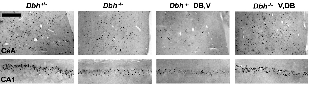
We previously described a role for adrenergic signaling in the hippocampus to promote contextual and spatial memory retrieval. A subsequent study performing expression analysis of the immediate-early gene (IEG) Arc suggested that activation of CA1 but not CA3 pyramidal neurons during memory retrieval is impaired in the absence of NE. The current study sought to confirm and extend those observations by performing expression analysis of a second IEG product, Fos, following a much greater variety of testing conditions. In mutant mice lacking NE, induction of Fos was normal in all regions of the hippocampus and amygdala shortly after fear conditioning. In contrast, when testing contextual fear 1 day after training, induction of Fos in CA1 and the central nucleus of the amygdala (CeA), but not CA3, the dentate gyrus or other amygdaloid nuclei, was impaired in the mutant mice. This pattern corresponded to the memory retrieval deficit exhibited by these mice. On the other hand, induction was normal in CA1 and CeA when testing cued fear 1 day after training, or contextual fear 1 week or 1 month after training, conditions in which retrieval are normal in the absence of NE. Acute restoration of NE in the mutant mice before testing but not before training rescued retrieval of contextual fear and restored Fos induction in CA1 and CeA. Because NE facilitates retrieval through the activation of β(1)-adrenergic receptors, β(1) knockout mice were also examined and found to exhibit reduced induction of Fos in CA1 and CeA following retrieval. Based on these and previous results, we hypothesize that adrenergic signaling is critical for the full activation of CA1 pyramidal neurons in response to excitatory input from CA3 pyramidal neurons conveying retrieved contextual information.


Figures

Fig. 1
Fig. 1
Coronal mouse brain diagram indicating regions where fos induction was quantified. For hippocampus: dDG, dCA3 and dCA1 are dorsal dentate gyrus, CA3 and CA1; and for amygdala: LA, BLA, BMA and CeA are lateral, basolateral, basomedial and central nuclei. The number in parentheses is the distance posterior to Bregma in mm.
Fig. 2
Fig. 2
Fos immunolabeling in DH and amygdala shortly after fear conditioning. Mice were sacrificed either unconditioned (U) or 2 h after conditioning (2). For all 7 brain regions (see Fig. 1), Fos labeling is significantly greater 2 h after training relative to no training (P < 0.001 for each main effect of conditioning). To examine the potential contribution of adrenergic signaling, mice with (Dbh+/−) and without (Dbh−/−) NE/E were studied. For all 7 brain regions, no significant differences between genotype were present (P > 0.4 for each main effect of genotype). For all figures, Fos-immunoreactive (Fos-IR) nuclei are normalized to 0.1 mm2 and n = 4–6 per group per genotype.
Fig. 3
Fig. 3
Fos immunolabeling in DH and amygdala following context reexposure one day after conditioning. (A) During reexposure to the conditioning apparatus, NE/E-deficient Dbh−/− mice froze significantly less than control Dbh+/− mice. (B) and (C) Mice were exposed to a neutral (non-shock) context (N) and the salient (shock) context (S) on training day. The next day, the mice were reexposed to one or the other context during testing and sacrificed 1 h later. Results were compared to those from unconditioned mice (U). For all 7 regions, Fos labeling is significantly greater in context S versus N, as indicated by the symbols with lines above the bars (P < 0.01 to 0.001 for each main effect of conditioning, as indicated). Significant differences in Fos labeling by genotype were present only in context S in CA1 and CeA. For CA1 and for CeA, P < 0.05 for the main effect of genotype and the interaction of condition and genotype. For all figures, significance is indicated as: *, P < 0.05; ^, P < 0.01; #, P < 0.001.
Fig. 4
Fig. 4
Representative Fos-immunolabeled sections from Dbh+/− and Dbh−/− mice following retrieval testing. The two panels on the left are from Dbh+/− and Dbh−/− mice that did not receive injections. Analogous results were obtained with Dbh+/− and Dbh−/− mice that received vehicle injections (V,V) 5 h prior to training and testing (Fig. 5). The two panels on the right are from Dbh−/− mice that received injections of L-DOPS with benserazide 5 h before training and vehicle 5 h before testing (DB,V), or vehicle 5 h before training and L-DOPS with benserazide 5 h before testing (V,DB). Scale bar is 50 µm.
Fig. 5
Fig. 5
Instatement of normal freezing and Fos labeling in Dbh−/− mice in response to the training context following restoration of NE before testing. Mice were injected 5 h before training and again 5 h before testing 2 days later with either vehicle (V) or L-DOPS plus benserazide (DB). Benserazide is a peripheral decarboxylase inhibitor that acts to restore NE selectively to the CNS in Dbh−/− mice. Only Dbh−/− mice treated with L-DOPS plus benserazide before testing (V,DB) exhibited freezing (A) and Fos labeling (B and C) that was comparable to that for Dbh+/− mice, consistent with the requirement for NE during contextual memory retrieval. For behavior, CA1 and CeA, P < 0.01 for the main effect of group.
Fig. 6
Fig. 6
Freezing and Fos labeling in Dbh−/− mice is normal following reexposure to the training context 7 or 31 days after fear conditioning. Mice were reexposed to the training context 1, 4, 7 or 31 days after training. Induction of Fos was significantly reduced in Dbh−/− mice at 1 and 4 days after training. In controls, there was a significant reduction in Fos labeling following reexposure to the training context at 31 days relative to earlier times in CA1 (P < 0.001), and at 7 and 31 days relative to earlier times in CeA (P < 0.05 for 7 days and P < 0.01 for 31 days). For behavior, P < 0.001 for the main effects of genotype and time, as well as the interaction of genotype and time. For CA1, P < 0.001 for the main effect of time, and P < 0.05 for the main effect of genotype and the interaction of genotype and time. For CeA, P < 0.001 for the main effect of time, P < 0.01 for the main effect of genotype, and P < 0.05 for the interaction of genotype and time.
Fig. 7
Fig. 7
Responses following cued fear testing one day after training. Mice were subjected to one of three conditions: reexposure to context N in the absence of the training tone (N); reexposure to context N and the training tone (N+T); reexposure to context N and the training tone following habituation to context N prior to conditioning (HN+T). No significant difference by genotype was present (P > 0.7 for each main effect of genotype). Note the dissociation between freezing and induction of Fos in CA1 but not CeA in the habituated (HN+T) mice. For behavior, CA1 and CeA, P < 0.001 for the main effect of condition.
Fig. 8
Fig. 8
Freezing and Fos induction are significantly reduced in β1 KO mice. Mice were reexposed to the training context one day after conditioning.

Similar articles

Cited by

References

    1. Anagnostaras SG, Gale GD, Fanselow MS. Hippocampus and contextual fear conditioning: recent controversies and advances. Hippocampus. 2001;11:8–17. - PubMed
    1. Booze RM, Crisostomo EA, Davis JN. Beta-adrenergic receptors in the hippocampal and retrohippocampal regions of rats and guinea pigs: autoradiographic and immunohistochemical studies. Synapse. 1993;13:206–214. - PubMed
    1. Cirelli C, Pompeiano M, Tononi G. Neuronal gene expression in the waking state: a role for the locus coeruleus. Science. 1996;274:1211–1215. - PubMed
    1. Cirelli C, Tononi G. Differential expression of plasticity-related genes in waking and sleep and their regulation by the noradrenergic system. J Neurosci. 2000;20:9187–9194. - PMC - PubMed
    1. Clayton DF. The genomic action potential. Neurobiol Learn Mem. 2000;74:185–216. - PubMed

Publication types

MeSH terms