. 2011 Jul;46(7):549-59.
doi: 10.1016/j.exger.2011.02.010. Epub 2011 Mar 3.

Cardiomyopathy of aging in the mammalian heart is characterized by myocardial hypertrophy, fibrosis and a predisposition towards cardiomyocyte apoptosis and autophagy

Affiliations

Cardiomyopathy of aging in the mammalian heart is characterized by myocardial hypertrophy, fibrosis and a predisposition towards cardiomyocyte apoptosis and autophagy

Andrew J Boyle et al. Exp Gerontol. 2011 Jul.

Abstract

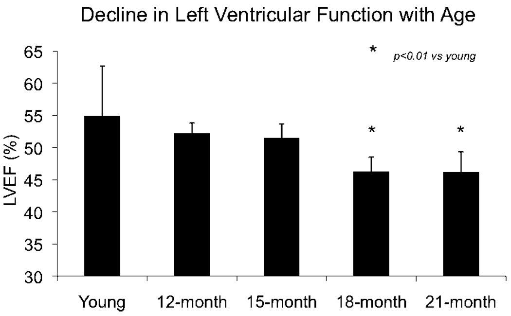
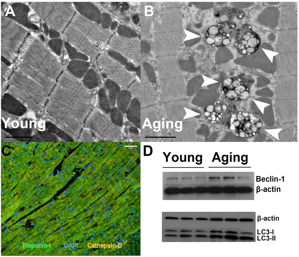
Aging is associated with an increased incidence of heart failure, but the existence of an age-related cardiomyopathy remains controversial. Differences in strain, age and technique of measuring cardiac function differ between experiments, confounding the interpretation of these studies. Additionally, the structural and genetic profile at the onset of heart failure has not been extensively studied. We therefore performed serial echocardiography, which allows repeated assessment of left ventricular (LV) function, on a cohort of the same mice every 3 months as they aged and demonstrated that LV systolic dysfunction becomes apparent at 18 months of age. These aging animals had left ventricular hypertrophy and fibrosis, but did not have inducible ventricular tachyarrhythmias. Gene expression profiling of left ventricular tissue demonstrated 40 differentially expressed probesets and 36 differentially expressed gene ontology terms, largely related to inflammation and immunity. At this early stage of cardiac dysfunction, we observed increased cardiomyocyte expression of the pro-apoptotic activated caspase-3, but no actual increase in apoptosis. The aging hearts also have higher levels of anti-apoptotic and autophagic factors, which may have rendered protection from apoptosis. In conclusion, we describe the functional, structural and genetic changes in murine hearts as they first develop cardiomyopathy of aging.


Figures

Figure 1
Figure 1
Serial echocardiography demonstrates a decline in left ventricular systolic function at 18 months of age.
Figure 2
Figure 2
Young hearts (A) demonstrate scant extracellular matrix (red color) on picrosirius red staining. Aging hearts (B) show expansion of extra cellular matrix. Polarized light microscopy (C young, D aging) shows that this is predominantly collagen, exhibiting birefringence. Quantification is shown below. Note that the increase in fibrosis and collagen is accompanied by cardiomyocyte hypertrophy, but there is no difference in blood pressure. Grey bars represent young, black bars represent aging. *p<0.01 vs young.
Figure 3
Figure 3
Aging hearts have higher cardiomyocyte expression of activated caspase-3 (A). Although activated caspase-3 can be seen in non-cardiomyocytes (troponin-I negative cells) in the young, there is little staining of cardiomyocytes. In the aged hearts, co-localization of troponin-I and activated caspase-3 (white arrows) indicates increased cardiomyocyte expression of active caspase-3. However, this does not translate into higher levels of cardiomyocyte apoptosis assessed by Hairpin-1 staining (B). Apoptotic cardiomyocytes are were rarely found (white arrow), and were not different between young and aging hearts. Caspase-independent mediators of apoptosis are not different in young and aging hearts (C). Characteristic cytoplasmic staining of AIF and BNIP-3, and peri-nuclear staining of EndoG are seen (white arrows). Anti-apoptotic protein expression appears higher in the aging heart (D). This suggests parallel activation of pro-apoptotic and pro-survival signals in aging cardiomyocytes, which results in no difference in the overall number of apoptotic cardiomyocytes. AIF = apoptosis inducing factor; EndoG = Endonuclease-G; BNIP3 = BCL2/adenovirus E1B 19 kDa protein-interacting protein 3; Bcl-2 = B-cell lymphoma 2; p-Akt = phosphorylated Akt.
Figure 4
Figure 4
Pro-apoptotic gene expression profile in aging hearts. Heatmap and gene list of apoptotic genes differentially expressed in young and aging hearts derived from geneset enrichment analysis. Red signifies increased expression, blue signifies reduced expression.
Figure 5
Figure 5
Activation of autophagy is seen in aging cardiomyocytes. (A,B) Electron microscopy demonstrates frequent autophagosomes in the aging heart (white arrowheads), but these are rare in the young heart. Autophagosomes are generally seen in close proximity to mitochondria. (D) Beclin-1 protein, a component of the autophagosome membrane, is upregulated in aging hearts. Aging heart demonstrate a shift from LC3 I to LC3 II, which is associated with induction of autophagy. Importantly, there is no blockage in autophagic flux to account for this finding, as demonstrated by similar levels of cathepsin D positive cardiomyocytes in young and aging hearts (C).
Figure 6
Figure 6
Inflammatory cells in the heart. A. Aging animals showed clusters of lymphocyte-like cells around blood vessels (green arrows) but young animals did not. B. Macrophages identified by CD68 staining were rarely seen in either the aging or young hearts. C. Mast cells, identified by toluidine blue staining (white arrow) were also rarely seen in either the aging or young hearts.

References

    1. NIH. National Heart Lung and Blood Institute Factbook Fiscal Year 2008
    1. Lloyd-Jones D, Adams R, Carnethon M, De Simone G, Ferguson TB, Flegal K, Ford E, Furie K, Go A, Greenlund K, Haase N, Hailpern S, Ho M, Howard V, Kissela B, Kittner S, Lackland D, Lisabeth L, Marelli A, McDermott M, Meigs J, Mozaffarian D, Nichol G, O'Donnell C, Roger V, Rosamond W, Sacco R, Sorlie P, Stafford R, Steinberger J, Thom T, Wasserthiel-Smoller S, Wong N, Wylie-Rosett J, Hong Y. Heart disease and stroke statistics--2009 update: a report from the American Heart Association Statistics Committee and Stroke Statistics Subcommittee. Circulation. 2009;119:480–486. - PubMed
    1. DHHS. A Profile of Older Americans: 2008
    1. Biesiadecki BJ, Tachampa K, Yuan C, Jin J-P, de Tombe PP, Solaro RJ. Removal of the Cardiac Troponin I N-terminal Extension Improves Cardiac Function in Aged Mice. Journal of Biological Chemistry. 2010;285:19688–19698. - PMC - PubMed
    1. Inuzuka Y, Okuda J, Kawashima T, Kato T, Niizuma S, Tamaki Y, Iwanaga Y, Yoshida Y, Kosugi R, Watanabe-Maeda K, Machida Y, Tsuji S, Aburatani H, Izumi T, Kita T, Shioi T. Suppression of Phosphoinositide 3-Kinase Prevents Cardiac Aging in Mice. Circulation. 2009;120:1695–1703. - PubMed

Publication types

MeSH terms