. 2011 Jun;300(6):L863-73.
doi: 10.1152/ajplung.00300.2009. Epub 2011 Mar 4.

A role for decorin in a murine model of allergen-induced asthma

Affiliations

A role for decorin in a murine model of allergen-induced asthma

Cinzia L Marchica et al. Am J Physiol Lung Cell Mol Physiol. 2011 Jun.

Abstract

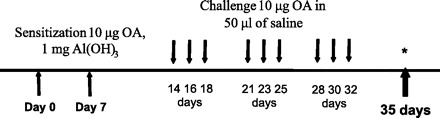
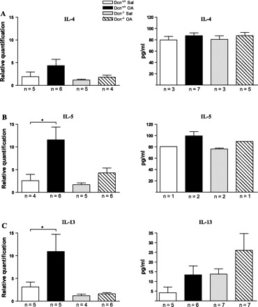
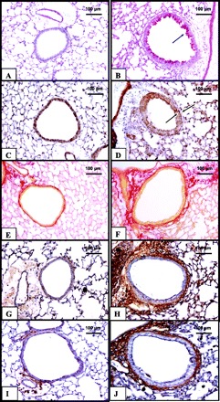
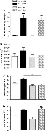
Decorin (Dcn) is an extracellular matrix proteoglycan, which affects airway mechanics, airway-parenchymal interdependence, airway smooth muscle proliferation and apoptosis, and transforming growth factor-β bioavailability. As Dcn deposition is differentially altered in asthma, we questioned whether Dcn deficiency would impact the development of allergen-induced asthma in a mouse model. Dcn(-/-) and Dcn(+/+) mice (C57Bl/6) were sensitized with ovalbumin (OA) and challenged intranasally 3 days/wk × 3 wk. After OA challenge, mice were anesthetized, and respiratory mechanics measured under baseline conditions and after delivery of increasing concentrations of methacholine aerosol. Complex impedance was partitioned into airway resistance and tissue elastance and damping. Bronchoalveolar lavage was performed. Lungs were excised, and tissue sections evaluated for inflammatory cell influx, α-smooth muscle actin, collagen, biglycan, and Dcn deposition. Changes in TH-2 cytokine mRNA and protein were also measured. Airway resistance was increased in OA-challenged Dcn(+/+) mice only (P < 0.05), whereas tissue elastance and damping were increased in both OA-challenged Dcn(+/+) and Dcn(-/-), but more so in Dcn(+/+) mice (P < 0.001). Inflammation and collagen staining within the airway wall were increased with OA in Dcn(+/+) only (P < 0.001 and P < 0.01, respectively, vs. saline). IL-5 and IL-13 mRNA were increased in lung tissue of OA-challenged Dcn(+/+) mice. Dcn deficiency resulted in more modest OA-induced hyperresponsiveness, evident at the level of the central airways and distal lung. Differences in physiology were accompanied by differences in inflammation and remodeling. These findings may be, in part, due to the well-described ability of Dcn to bind transforming growth factor-β and render it less bioavailable.


Figures

Fig. 1.
Fig. 1.
Antigen sensitization and challenge protocol. Experimental groups were sensitized on two separate occasions, 1 wk apart, with ovalbumin (OA) and adjuvant aluminum hydroxide. This was followed by a total of 9 challenges by intranasal instillation of OA in saline (Sal; 3 days/wk for 3 wk). Physiological assessments were carried out on day 35. Control mice were sensitized and challenged with Sal alone.
Fig. 2.
Fig. 2.
Lung physiology measurements evaluating changes in central airway resistance (Raw; A), tissue damping (Gti; B), and tissue elastance (Hti; C) during methacholine (MCh) aerosol challenges in OA-challenged and Sal control decorin (Dcn)+/+ and Dcn−/− mice. A: Raw was significantly increased in the Dcn+/+ OA group only, compared with Sal-challenged mice. Moreover, the Raw response observed in OA Dcn−/− mice was significantly lower than that of OA-challenged Dcn+/+ mice at MCh dose of 25 mg/ml. B and C: Gti and Hti responses were significantly increased in Dcn+/+ as well as Dcn−/− OA-exposed mice compared with Sal, although this response was apparent at a lower dose of MCh (25 mg/ml) in the Dcn+/+ group. In addition, Gti and Hti responses were significantly higher in Dcn+/+ then in Dcn−/− OA groups at MCh of 25 and 50 mg/ml. Values are means ± SE. *P < 0.05, OA vs. Sal. ***P < 0.001, OA vs. Sal. †P < 0.05, Dcn−/− vs. Dcn+/+. ††P < 0.01, Dcn−/− vs. Dcn+/+. †††P < 0.001, Dcn−/− vs. Dcn+/+.
Fig. 3.
Fig. 3.
AD: photomicrographs depicting tissue inflammation (hematoxylin and eosin). Magnification ×100 μm. A: Dcn+/+ Sal control. B: Dcn+/+ OA challenged. C: Dcn−/− Sal control. D: Dcn−/− OA challenged. Tissue inflammation, determined by the presence of inflammatory cells (as displayed by the arrow), is present in greater amounts in the OA-challenged Dcn+/+ group (B) compared with Sal control (A). Furthermore, no significant inflammation is seen with OA in Dcn−/− (D). E: tissue inflammation quantified in the airway wall as the number of inflammatory cells/wall area (WA) (mm2). Tissue inflammation was shown to be significantly increased in OA-challenged Dcn+/+ mice only, compared with Dcn+/+ Sal mice (***P < 0.001). The increase observed in OA-exposed Dcn+/+ mice was, in addition, significantly higher than the level of tissue inflammation present in the OA Dcn−/− group (†††P < 0.001). F: bronchoalveolar lavage (BAL) cell differential. Percentage of eosinophils, macrophages, neutrophils, and lymphocytes determined out of a total of 300 cells counted. OA-challenged mice demonstrated significant increases in the percentage of eosinophil (***P < 0.001), along with concomitant decreases in the percentage of macrophages (***P < 0.001). No significant changes were observed in the percentage of neutrophils or lymphocytes across all four groups. Values are means ± SE.
Fig. 4.
Fig. 4.
TH-2 cytokines. Levels of TH-2 cytokines, IL-4 (A), IL-5 (B), and IL-13 (C), of mRNA message were evaluated in lung tissue using quantitative PCR (left), as well as protein levels in BAL, as quantified using ELISA (right). IL-4 mRNA expression (A, left) was not found to be significantly different between the experimental groups. IL-5 (B, left) and IL-13 (C, left) were significantly increased in the lung tissue of OA-challenged Dcn+/+ mice compared with Sal-challenged Dcn+/+ mice (*P < 0.05). The OA challenged did not induce an increase in the mRNA expression of these TH-2 cytokine Dcn−/− mice. IL-4 (A, right) and IL-5 (B, right) BAL levels were not significantly different among groups. C: IL-13 was not significantly different among the four experimental groups. Values are means ± SE; n, no. of animals.
Fig. 5.
Fig. 5.
Representative photomicrographs of histochemical and immunohistochemical staining in the airway wall of Sal-challenged Dcn+/+ (C, E, and I) and Dcn−/− (A and G), as well as OA-challenged Dcn+/+ (D, F, and J) and Dcn−/− (B and H) mice. Staining is shown of airways with periodic acid Schiff (PAS) (A and B), primary antibody for α-smooth muscle actin (α-SMA; C and D), picrosirius red (E and F), primary antibody for biglycan (Bgn) (G and H), and primary antibody for Dcn (I and J). Arrows in B and D identify goblets cells positive for mucus expression and smooth muscle (SM) bundles, respectively. Photomicrographs (E and F) reveal positive staining for collagen fibers shown in red, present around the airways. Positive staining for Bgn, displayed in brown, shows increased Bgn deposition with OA challenge in Dcn−/− mice. Dcn-positive stain, also shown in brown, is demonstrated to be increased with OA challenge in Dcn+/+ mice. Magnification ×100 μm.
Fig. 6.
Fig. 6.
Quantification of airway mucous (PAS), α-SMA, collagen, and Bgn immunostaining. A: mucus expression was significantly increased with OA in both Dcn+/+ and Dcn−/− mice (***P < 0.001). B: Dcn+/+ Sal, Dcn−/− Sal, and Dcn−/− OA-exposed mice demonstrated approximately the same amount of positive staining for α-SMA, whereas OA-challenged Dcn+/+ mice exhibited a slightly increased amount of α-SMA staining. C: OA challenge significantly increased collagen deposition around the airways of Dcn+/+ mice only, compared with Dcn+/+ Sal controls (*P < 0.05). Collagen deposition was not observed to be altered between OA and Sal Dcn−/− mice. In addition, the amount of collagen deposition present in OA Dcn+/+ mice was significantly higher than that present in OA Dcn−/− mice (††P < 0.01). D: increased Bgn deposition was observed with OA challenge in both Dcn+/+ and Dcn−/− mice compared with their respective Sal controls. This increase, however, was found to be statistically significant when comparing OA vs. Sal Dcn−/− mice only (***P < 0.001). Values are means ± SE. Pbm2, basement membrane perimeter squared.
Fig. 7.
Fig. 7.
Transforming growth factor (TGF-β) levels. A: total lung proteins were extracted and analyzed by Western blot using a specific antibody for TGF-β1. B: densitometric analysis of Western blot corrected for β-actin levels shows increases in TGF-β1 in OA-challenged mice vs. Sal controls, although this did not reach statistical significance. C: BAL levels of TGF-β1, using ELISA, were decreased in the OA-challenged Dcn−/− mice compared with OA-challenged Dcn+/+ mice (***P < 0.001). D: TGF-β1 mRNA expression in extracted lung tissue, as quantified using quantitative PCR, was not found to be significantly different among the experimental groups. Values are means ± SE.

Similar articles

Cited by

References

    1. Al JR, Roughley PJ, Ludwig MS. Effect of glycosaminoglycan degradation on lung tissue viscoelasticity. Am J Physiol Lung Cell Mol Physiol 280: L306–L315, 2001. - PubMed
    1. Alcorn JF, Rinaldi LM, Jaffe EF, van LM, Bates JH, Janssen-Heininger YM, Irvin CG. Transforming growth factor-beta1 suppresses airway hyperresponsiveness in allergic airway disease. Am J Respir Crit Care Med 176: 974–982, 2007. - PMC - PubMed
    1. Busse WW. Leukotrienes and inflammation. Am J Respir Crit Care Med 157: S210–S213, 1998. - PubMed
    1. Cavalcante FS, Ito S, Brewer K, Sakai H, Alencar AM, Almeida MP, Andrade JS, Jr, Majumdar A, Ingenito EP, Suki B. Mechanical interactions between collagen and proteoglycans: implications for the stability of lung tissue. J Appl Physiol 98: 672–679, 2005. - PubMed
    1. D'Antoni ML, Torregiani C, Ferraro P, Michoud MC, Mazer B, Martin JG, Ludwig MS. Effects of decorin and biglycan on human airway smooth muscle cell proliferation and apoptosis. Am J Physiol Lung Cell Mol Physiol 294: L764–L771, 2008. - PubMed

Publication types

MeSH terms