. 2011 Apr 22;286(16):14246-56.
doi: 10.1074/jbc.M110.147348. Epub 2011 Mar 4.

7,3',4'-Trihydroxyisoflavone, a metabolite of the soy isoflavone daidzein, suppresses ultraviolet B-induced skin cancer by targeting Cot and MKK4

Affiliations

7,3',4'-Trihydroxyisoflavone, a metabolite of the soy isoflavone daidzein, suppresses ultraviolet B-induced skin cancer by targeting Cot and MKK4

Dong Eun Lee et al. J Biol Chem. .

Abstract

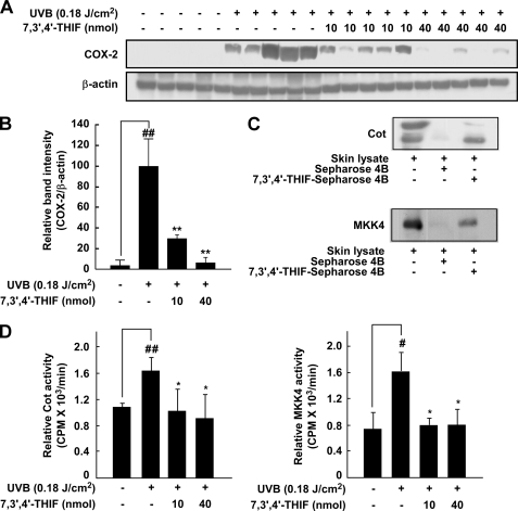
Nonmelanoma skin cancer is one of the most frequently occurring cancers in the United States. Chronic exposure to UVB irradiation is a major cause of this cancer. Daidzein, along with genistein, is a major isoflavone found in soybeans; however, little is known about the chemopreventive effects of daidzein and its metabolites in UVB-induced skin cancer. Here, we found that 7,3',4'-trihydroxyisoflavone (THIF), a major metabolite of daidzein, effectively inhibits UVB-induced cyclooxygenase 2 (COX-2) expression through the inhibition of NF-κB transcription activity in mouse skin epidermal JB6 P+ cells. In contrast, daidzein had no effect on COX-2 expression levels. Data from Western blot and kinase assays showed that 7,3',4'-THIF inhibited Cot and MKK4 activity, thereby suppressing UVB-induced phosphorylation of mitogen-activated protein kinases. Pull-down assays indicated that 7,3',4'-THIF competed with ATP to inhibit Cot or MKK4 activity. Topical application of 7,3',4'-THIF clearly suppressed the incidence and multiplicity of UVB-induced tumors in hairless mouse skin. Hairless mouse skin results also showed that 7,3',4'-THIF inhibits Cot or MKK4 kinase activity directly, resulting in suppressed UVB-induced COX-2 expression. A docking study revealed that 7,3',4'-THIF, but not daidzein, easily docked to the ATP binding site of Cot and MKK4, which is located between the N- and C-lobes of the kinase domain. Collectively, these results provide insight into the biological actions of 7,3',4'-THIF, a potential skin cancer chemopreventive agent.


Figures

FIGURE 1.
FIGURE 1.
Comparison of the inhibitory effects of 7,3′,4′-THIF and daidzein on UVB-induced COX-2 expression, COX-2 promoter activity, and NF-κB transactivation in JB6 P+ cells. A, the chemical structures of 7,3′,4′-THIF and daidzein. B, effect of 7,3′,4′-THIF or daidzein on UVB-induced COX-2 expression in JB6 P+ cells. The cells were treated with 7,3′,4′-THIF or daidzein at the indicated concentrations for 1 h before exposure to UVB (4 kJ/m2) and harvested 4 h later. The levels of COX-2 expression were then determined by Western blot analysis, as described under “Materials and Methods,” using specific antibodies against the corresponding COX-2 and β-actin proteins. Data are representative of three independent experiments. The protein levels were quantified using an image analysis program to evaluate the density of each band on the immunoblot, and the fold value was calculated. C and D, effects of 7,3′,4′-THIF or daidzein on UVB-induced COX-2 (C) or NF-κB (D) activity in JB6 P+ cells. In the luciferase assay, JB6 P+ stably transfected with the COX-2 or NF-κB luciferase reporter plasmid were cultured as described under “Materials and Methods.” After reaching 80% confluence, the cells were cultured in 0.1% FBS/MEM and then treated with 7,3′,4′-THIF or daidzein at the indicated concentrations or left untreated for 1 h before exposure to UVB (4 kJ/m2) and harvested 24 h later. COX-2 or NF-κB activity is expressed as the percent inhibition relative to cells treated with only UVB. Data are represented as the mean ± S.D. of the luciferase activity calculated from three separate experiments. For C and D, the asterisk indicates significant differences between groups treated with UVB alone and the 7,3′,4′-THIF- or daidzein-treated groups. *, p < 0.05; **, p < 0.01; ***, p < 0.001).
FIGURE 2.
FIGURE 2.
7,3′,4′-THIF suppresses UVB-induced phosphorylation of JNKs and p38 through the direct inhibition of both Cot and MKK4. A, 7,3′,4′-THIF inhibited UVB-induced phosphorylation of JNKs and p38 in JB6 P+ cells. Cells were treated with 7,3′,4′-THIF (0, 20, 40, or 60 μm) for 1 h before being exposed to UVB (4 kJ/m2) and harvested 30 min later. The cells were disrupted, and the levels of phosphorylated and total proteins were determined by Western blot analysis, as described under “Materials and Methods,” using specific antibodies against the respective phosphorylated and total proteins. Data are representative of three independent experiments that gave similar results. B, 7,3′,4′-THIF inhibited both Cot and MKK4 in vitro. In contrast, MKK3 and MKK6 activity was not affected by 7,3′,4′-THIF. The in vitro kinase assay was performed as described under “Materials and Methods,” and kinase activity is expressed as percent inhibition relative to the activity of the untreated kinase control. C, 7,3′,4′-THIF inhibited both Cot and MKK4 activity ex vivo. In the ex vivo Cot or MKK4 kinase assay, cells were pretreated with 7,3′,4′-THIF at the indicated concentrations (0, 20, 40, or 60 μm) for 1 h and then exposed to UVB (4 kJ/m2) and harvested after 30 min. Cells were used for immunoprecipitation, and the kinase assay was performed. Kinase activity is expressed as percent inhibition relative to cells treated with UVB only. The average 32P count was determined from three separate experiments, and the data are presented as mean ± S.D. In the ex vivo kinase assays, the asterisks indicate a significant decrease in kinase activity between cells treated with 7,3′,4′-THIF and cells treated with UVB only. **, p < 0.01; ***, p < 0.001). D, 7,3′,4′-THIF did not affect ERKs or MSK1 activity in vitro. The in vitro kinase assay was performed as described under “Materials and Methods,” and kinase activity is expressed as percent inhibition relative to the activity of the untreated kinase control. For the in vitro kinase assays (B and D), the asterisk indicates a significant decrease in kinase activity between the groups treated with active Cot (or MKK4) and 7,3′,4′,-THIF and the group treated with active Cot (or MKK4) alone. *, p < 0.05; **, p < 0.01; ***, p < 0.001.
FIGURE 3.
FIGURE 3.
7,3′,4′-THIF direct-binds with Cot and MKK4. A, 7,3′,4′-THIF specifically binds with Cot or MKK4 in vitro. The Cot (or MKK4) 7,3′,4′-THIF binding was confirmed by immunoblotting using an antibody against Cot (left panel) or MKK4 (right panel). 1st lane (input control), Cot, or MKK4 protein standard; 2nd lane (control), Sepharose 4B was used to pull down Cot or MKK4, as described under “Materials and Methods;” 3rd lane, 7,3′,4′-THIF-Sepharose 4B affinity beads were used to pull down Cot or MKK4. B, 7,3′,4′-THIF directly binds with either Cot and MKK4 ex vivo. Binding was confirmed by immunoblotting using an antibody against Cot (left panel) or MKK4 (right panel). 1st lane (input control), whole cell lysates from JB6 P+ cells; 2nd lane (control), a lysate of JB6 P+ cells precipitated with Sepharose 4B beads; 3rd lane, whole cell lysates from JB6 P+ cells precipitated by 7,3′,4′-THIF-Sepharose 4B affinity beads. C, 7,3′,4′-THIF competes with ATP to bind with Cot and MKK4. Active Cot or MKK4 (0.2 μg) was incubated with ATP at different concentrations (0, 10, or 100 μm) and 100 μl of 7,3′,4′-THIF-Sepharose 4B or 100 μl of Sepharose 4B (as a negative control) in reaction buffer to a final volume of 500 μl. The mixtures were incubated at 4 °C overnight with shaking. After washing, the pulled-down proteins were detected by Western blotting. 1st lane (input control), Cot or MKK4 protein standard; 2nd lane, (negative control), Cot or MKK4 bound with Sepharose 4B; 3rd lane (positive control), Cot (left panel) or MKK4 (right panel) binding with 7,3′,4′-THIF-Sepharose 4B. Each experiment was performed three times, and representative blots are shown.
FIGURE 4.
FIGURE 4.
7,3′,4′-THIF inhibits UVB-induced skin carcinogenesis in the SKH-1 hairless mouse. Control mice (n = 12) received a topical treatment of 200 μl acetone (no UVB), and experimental mice (n = 12) were topically treated with 200 μl acetone before UVB (0.18 J/cm2) exposure (3 days/week for 27 weeks). The mice in the third and fourth groups received a topical application of 7,3′,4′-THIF (10 or 40 nmol, respectively, per mouse in 200 μl acetone) on the dorsal surface 1 h before UVB (0.18 J/cm2) irradiation 3 days/week for 27 weeks. The incidence of skin tumors was recorded weekly, and tumors were defined as an outgrowth of >1 mm in diameter persisting for 2 weeks or longer. Tumor incidence and multiplicity were recorded every week until the end of the experiment at 27 weeks. A, appearance of skin tumors. B, 7,3′,4′-THIF retarded the incidence of skin tumors compared with the UVB only-treated group. C, 7,3′,4′-THIF strongly inhibits UVB-induced tumor multiplicity in SKH-1 hairless mice. D, 7,3′,4′-THIF reduces UVB-induced tumor volume in SKH-1 hairless mice. At the end of the study, the dimensions of all tumors on each mouse were recorded. Tumor volumes were calculated using the hemiellipsoid model formula: tumor volume = 1/2(4π/3)(l/2)(w/2)h, wherein l is length, w is width, and h is height. The asterisks indicate a significant decrease in tumor incidence or volume between the group treated with UVB alone and the group treated with 7,3′,4′-THIF. **, p < 0.05; ***, p < 0.001.
FIGURE 5.
FIGURE 5.
7,3′,4′-THIF suppresses UVB-induced COX-2 expression and Cot and MKK4 kinase activity in the SKH-1 hairless mouse. A, 7,3′,4′-THIF inhibits UVB-induced COX-2 expression in SKH-1 hairless mouse skin. Five mouse skin samples were randomly selected from 12 mouse skin samples of each group and analyzed for COX-2 expression by immunoblotting. B, COX-2 immunoblot results were normalized to β-actin followed by statistical analysis of relative image density. ##, significant difference (p < 0.01) between the control group and the untreated group; *, significant difference at p < 0.05 between 7,3′,4′-THIF-treated groups and untreated groups. C, 7,3′,4′-THIF directly binds with either Cot or MKK4 in SKH-1 hairless mouse skin. Cot or MKK4–7,3′,4′-THIF binding was confirmed by immunoblotting using an antibody against Cot (upper panel) or MKK4 (lower panel). 1st lane (input control), whole skin lysates from SKH-1 hairless mice; 2nd lane (control), a lysate of mouse skin precipitated with Sepharose 4B beads; 3rd lane, whole skin lysates from SKH-1 hairless mice precipitated by 7,3′,4′-THIF-Sepharose 4B beads. D, 7,3′,4′-THIF inhibits UVB-induced Cot or MKK4 kinase activity in hairless mouse skin extracts. In the Cot (or MKK4) kinase assay, dorsal skin protein lysates were prepared from the epidermis, and immunoprecipitation and kinase assays were performed as described under “Materials and Methods.” Columns, mean of the 32P count from three separate experiments; bars, S.D., #, significant difference (p < 0.05); ##, significant difference (p < 0.001) between the control group and the group exposed to UVB; *, significant difference at p < 0.05 between groups treated with 7,3′,4′-THIF and untreated groups.
FIGURE 6.
FIGURE 6.
Modeling study of Cot or MKK4 binding with 7,3′,4′-THIF. A, hypothetical model of the Cot-7,3′,4′-THIF complex. 7,3′,4′-THIF (atomic color with green carbon) binds to the ATP-binding site in the kinase domain of Cot. The residues involved in the interactions with 7,3′,4′-THIF are indicated. The hydrogen bond is depicted as a white line. B, hypothetical model of the MKK4–7,3′,4′-THIF complex. 7,3′,4′-THIF (atomic color with green carbon) binds to the ATP-binding site in the kinase domain of MKK4. The hydrogen bond is depicted as a white line. C, Hypothetical model of Cot with bound ATP (atomic color with green carbon). D, hypothetical model of MKK4 with bound ATP (atomic color with green carbon).

References

    1. Athar M., An K. P., Morel K. D., Kim A. L., Aszterbaum M., Longley J., Epstein E. H., Jr., Bickers D. R. (2001) Biochem. Biophys. Res. Commun. 280, 1042–1047 - PubMed
    1. Rundhaug J. E., Fischer S. M. (2008) Photochem. Photobiol. 84, 322–329 - PubMed
    1. Katiyar S. K., Korman N. J., Mukhtar H., Agarwal R. (1997) J. Natl. Cancer Inst. 89, 556–566 - PubMed
    1. Buckman S. Y., Gresham A., Hale P., Hruza G., Anast J., Masferrer J., Pentland A. P. (1998) Carcinogenesis 19, 723–729 - PubMed
    1. Marwaha V., Chen Y. H., Helms E., Arad S., Inoue H., Bord E., Kishore R., Sarkissian R. D., Gilchrest B. A., Goukassian D. A. (2005) J. Biol. Chem. 280, 32379–32388 - PubMed

Publication types

MeSH terms