. 2011 Apr;60(4):1111-21.
doi: 10.2337/db10-1178. Epub 2011 Mar 4.

Bimodal effect on pancreatic β-cells of secretory products from normal or insulin-resistant human skeletal muscle

Affiliations

Bimodal effect on pancreatic β-cells of secretory products from normal or insulin-resistant human skeletal muscle

Karim Bouzakri et al. Diabetes. 2011 Apr.

Erratum in

  • Diabetes. 2015 Jan;64(1):312

Abstract

Objective: Type 2 diabetes is characterized by insulin resistance with a relative deficiency in insulin secretion. This study explored the potential communication between insulin-resistant human skeletal muscle and primary (human and rat) β-cells.

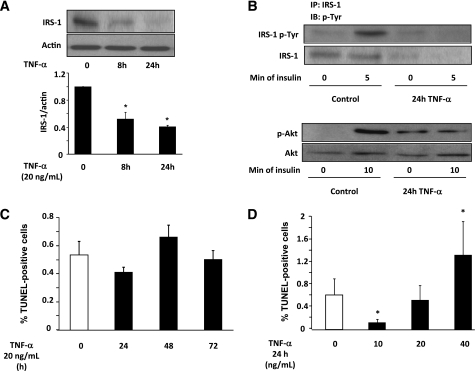
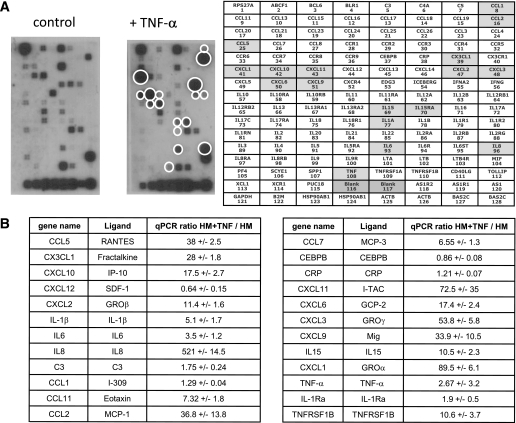
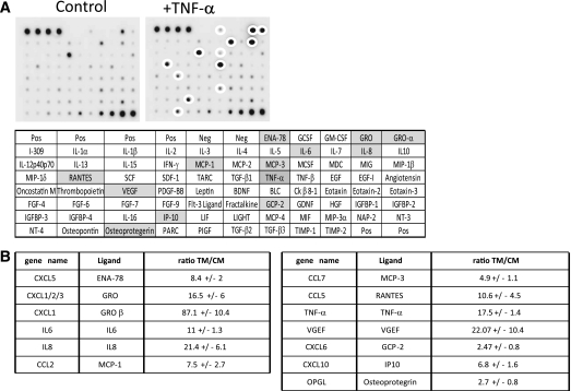
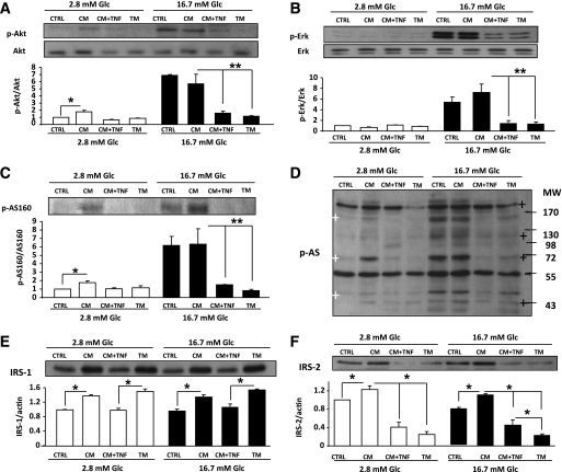
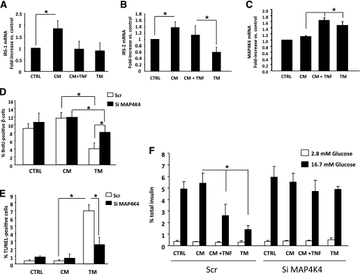
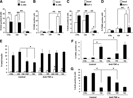
Research design and methods: Human skeletal muscle cells were cultured for up to 24 h with tumor necrosis factor (TNF)-α to induce insulin resistance, and mRNA expression for cytokines was analyzed and compared with controls (without TNF-α). Conditioned media were collected and candidate cytokines were measured by antibody array. Human and rat primary β-cells were used to explore the impact of exposure to conditioned media for 24 h on apoptosis, proliferation, short-term insulin secretion, and key signaling protein phosphorylation and expression.

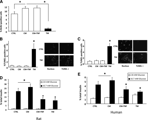
Results: Human myotubes express and release a different panel of myokines depending on their insulin sensitivity, with each panel exerting differential effects on β-cells. Conditioned medium from control myotubes increased proliferation and glucose-stimulated insulin secretion (GSIS) from primary β-cells, whereas conditioned medium from TNF-α-treated insulin-resistant myotubes (TMs) exerted detrimental effects that were either independent (increased apoptosis and decreased proliferation) or dependent on the presence of TNF-α in TM (blunted GSIS). Knockdown of β-cell mitogen-activated protein 4 kinase 4 prevented these effects. Glucagon-like peptide 1 protected β-cells against decreased proliferation and apoptosis evoked by TMs, while interleukin-1 receptor antagonist only prevented the latter.

Conclusions: Taken together, these data suggest a possible new route of communication between skeletal muscle and β-cells that is modulated by insulin resistance and could contribute to normal β-cell functional mass in healthy subjects, as well as the decrease seen in type 2 diabetes.


Figures

FIG. 1.
FIG. 1.
Effect of TNF-α on insulin sensitivity and apoptosis in human skeletal muscle cells. A: Human myotubes were cultured 24 h ± 20 ng/mL TNF-α, and IRS-1 protein expression was measured by Western blot (normalized to actin). Mean ± SE, 5 independent experiments. B: 24 h after incubation with TNF-α, human skeletal muscle cells were treated with 100 nmol/L insulin either for 5 min to induce IRS-1 tyrosine phosphorylation or 10 min to measure Akt phosphorylation. Representative immunoblots are shown for IRS-1 tyrosine (Tyr) phosphorylation (upper panel) and Akt Ser479 phosphorylation (lower panel). C: Effect of different times of TNF-α treatment (20 ng/mL) on human skeletal muscle cell death measured by TUNEL assay. n = 5 independent experiments. D: Effect of different TNF-α concentrations (24 h) on human primary skeletal muscle cell death measured by TUNEL assay. n = 5 independent experiments; *P < 0.05 vs. control. IP, immunoprecipitation; IB, immunoblotting.
FIG. 2.
FIG. 2.
Effect of TNF-α on expression of cytokine genes in human skeletal muscle cells. A: Human myotubes were cultured 8 h ± 20 ng/mL TNF-α, and cytokine mRNA levels were monitored by hybridization to Oligo nucleotide array membranes. White circles show modified mRNA levels between the two conditions on representative membranes out of four different experiments. B: Cytokine mRNA levels were measured by quantitative RT-PCR (qRT-PCR). The data are the mean value of four independent experiments, and mRNA expression levels were normalized to cyclophilin A. HM + TNF, human skeletal muscle cells after 8-h culture with TNF-α; HM, human skeletal muscle cells after 8-h culture without TNF-α.
FIG. 3.
FIG. 3.
Effect of TNF-α on human skeletal muscle cell cytokine secretion. A: Human myotubes were cultured 24 h ± 20 ng/mL TNF-α, and cytokine levels in the conditioned media were monitored by hybridization to human chemokine + cytokine array membranes. White circles show modified protein levels between the two conditions on representative membranes from three different experiments. B: Each membrane was quantified using Multi Gauge software, and the intensity was normalized to internal positive controls for comparison. n = 3 independent experiments. TM, medium from human skeletal muscle cells cultured 24 h with TNF-α; CM, medium from human skeletal muscle cells cultured 24 h without TNF-α.
FIG. 4.
FIG. 4.
Effect of conditioned medium on primary β-cell proliferation, survival, and GSIS. Conditioned medium was obtained by culturing human myotubes for 24 h with (TM) or without (CM) 20 ng/mL TNF-α. Additional control conditions were as follows: CTRL, culture medium not previously exposed to skeletal muscle cells; CM + TNF, 20 ng/mL TNF-α added to CM immediately before exposure to β-cells. A: Proliferation of rat primary β-cells measured by BrdU incorporation. Cells were grown under standard culture conditions (20% FCS, 11.2 mmol/L glucose) and treated for 48 h with the different conditioned media; BrdU was added for the last 24 h. β-Cells were identified by insulin immunofluorescence. n = 7 independent experiments. B and C: Rat and human primary β-cell apoptosis. Cell death was measured by TUNEL. n = 7 (rats) and n = 5 (human) independent experiments. *P < 0.05. D and E: Glucose-stimulated insulin secretion from rat and human primary β-cells measured during 60 min at 2.8 mmol/L glucose (white bars = basal secretion) following by 60 min at 16.7 mmol/L glucose (dark bars = stimulated secretion). Secretion is expressed as a percentage total insulin content. n = 7 (rats) and n = 5 (human) independent experiments. *P < 0.05.
FIG. 5.
FIG. 5.
Action of conditioned medium on Akt, ERK, AS160, and Akt substrates phosphorylation after glucose stimulation, or IRS-1 and -2 protein expression in rat primary β-cells. Conditioned media and abbreviations as described in the legend to Fig. 3. After 24-h culture in conditioned medium, β-cells were incubated 1 h at 2.8 mmol/L or 16.7 mmol/L glucose (Glc). Open bars = 2.8 mmol/L glucose; closed bars = 16.7 mmol/L glucose. Western blots were scanned and data normalized to total protein or actin as indicated. N = 3 independent experiments. A: Akt Ser 473 phosphorylation. *P < 0.05; **P < 0.01. B: ERK1/2 phosphorylation. **P < 0.01. C: AS160 phosphorylation. *P < 0.05; **P < 0.01. D: Ser/Thr phosphorylation of Akt substrates (white crosses = CM effect; black crosses = TM effect). E and F: IRS-1 (E) and IRS-2 (F) protein expression. *P < 0.05. CTRL, control.
FIG. 6.
FIG. 6.
Action of conditioned medium on IRS-1, IRS-2, and MAP4K4 mRNA expression and effect of MAP4K4 knockdown. Conditioned media and abbreviations as described in the legend to Fig. 3. Rat primary β-cells were used for n = 5 independent experiments. AC: IRS-1, IRS-2, and MAP4K4 mRNA expression were measured by quantitative real-time RT-PCR in β-cells. *P < 0.05. D–F: Proliferation (BrdU incorporation) (D) and apoptosis (TUNEL) (E) were measured in β-cells transfected with scrambled (Scr) (open bars = Scr) or with MAP4K4 small interfering RNA (siRNA) Si MAP4K4 (closed bars = Si MAP4K4). F: Insulin secretion was measured using β-cells transfected with scrambled (Scr) or with Si MAP4K4 and incubated for 60 min at 2.8 mmol/L glucose (open bars) followed by 60 min at 16.7 mmol/L glucose (closed bars). *P < 0.05. CTRL, control.
FIG. 7.
FIG. 7.
Effect of IL-1RA, GLP-1, or anti–TNF-α on rat primary β-cells exposed to conditioned medium. Conditioned media and abbreviations as described in the legend to Fig. 3. Rat primary β-cells were treated 48 h with IL-1RA (1 μg/mL) and conditioned medium. A: BrdU was added during the last 24 h to measure proliferation. B: Conditioned medium added for the last 24 h and cell death was measured by TUNEL. C: Rat primary β-cells were treated 48 h with conditioned medium, with or without GLP-1 (100 nmol/L), and BrdU was added the last 24 h to measure proliferation. D: Rat primary β-cells were treated 48 h with or without GLP-1, and conditioned medium was added for the last 24 h. Cell death was measured as described. n = 3 independent experiments. *P < 0.05; **P < 0.01. E: Insulin secretion was measured using rat primary β-cells treated for 24 h with the different conditioned media with or without TNF-α blockade (2 ng/mL Ethanercept) and then incubated for 60 min at 2.8 mmol/L glucose (open bars) followed by 60 min at 16.7 mmol/L glucose (closed bars); *P < 0.05. F and G: Rat primary β-cells treated with the different conditioned media with or without Ethanercept and were used to assess cell death using TUNEL assay (F) and cell proliferation (G) as described. n = 4 independent experiments; *P < 0.05 for TNF-α blockade. CTRL, control.

References

    1. Bluestone JA, Herold K, Eisenbarth G. Genetics, pathogenesis and clinical interventions in type 1 diabetes. Nature 2010;464:1293–130010.1038/nature08933 - DOI - PMC - PubMed
    1. Butler AE, Janson J, Bonner-Weir S, Ritzel R, Rizza RA, Butler PC. Beta-cell deficit and increased beta-cell apoptosis in humans with type 2 diabetes. Diabetes 2003;52:102–11010.2337/diabetes.52.1.102 - DOI - PubMed
    1. Wentworth JM, Fourlanos S, Harrison LC. Reappraising the stereotypes of diabetes in the modern diabetogenic environment. Nat Rev Endocrinol 2009;5:483–48910.1038/nrendo.2009.149 - DOI - PubMed
    1. Pedersen BK, Steensberg A, Fischer C, et al. The metabolic role of IL-6 produced during exercise: is IL-6 an exercise factor? Proc Nutr Soc 2004;63:263–26710.1079/PNS2004338 - DOI - PubMed
    1. Ellingsgaard H, Ehses JA, Hammar EB, et al. Interleukin-6 regulates pancreatic alpha-cell mass expansion. Proc Natl Acad Sci USA 2008;105:13163–1316810.1073/pnas.0801059105 - DOI - PMC - PubMed

Publication types

MeSH terms