. 2011 May;193(9):2290-300.
doi: 10.1128/JB.01555-10. Epub 2011 Mar 4.

Characterization of mutants deficient in the L,D-carboxypeptidase (DacB) and WalRK (VicRK) regulon, involved in peptidoglycan maturation of Streptococcus pneumoniae serotype 2 strain D39

Affiliations

Characterization of mutants deficient in the L,D-carboxypeptidase (DacB) and WalRK (VicRK) regulon, involved in peptidoglycan maturation of Streptococcus pneumoniae serotype 2 strain D39

Skye M Barendt et al. J Bacteriol. 2011 May.

Abstract

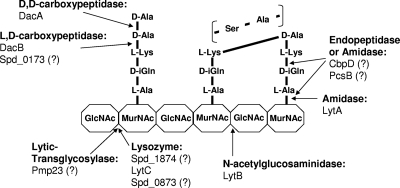
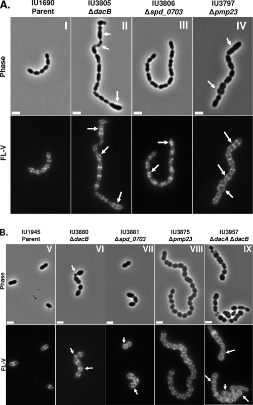
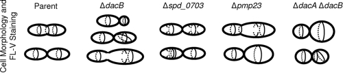
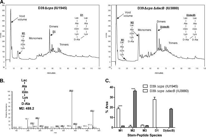
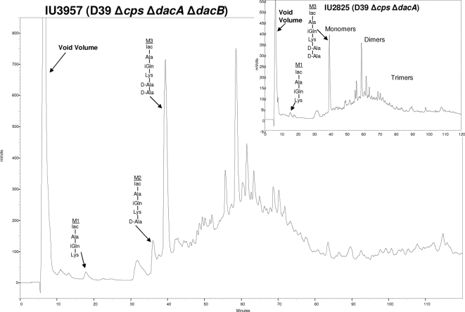
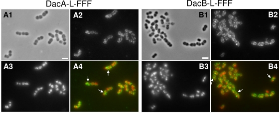
Peptidoglycan (PG) hydrolases play critical roles in the remodeling of bacterial cell walls during division. PG hydrolases have been studied extensively in several bacillus species, such as Escherichia coli and Bacillus subtilis, but remain relatively uncharacterized in ovococcus species, such as Streptococcus pneumoniae (pneumococcus). In this work, we identified genes that encode proteins with putative PG hydrolytic domains in the genome of S. pneumoniae strain D39. Knockout mutations in these genes were constructed, and the resulting mutants were characterized in comparison with the parent strain for growth, cell morphology, PG peptide incorporation, and in some cases, PG peptide composition. In addition, we characterized deletion mutations in nonessential genes of unknown function in the WalRK(Spn) two-component system regulon, which also contains the essential pcsB cell division gene. Several mutants did not show overt phenotypes, which is perhaps indicative of redundancy. In contrast, two new mutants showed distinct defects in PG biosynthesis. One mutation was in a gene designated dacB (spd_0549), which we showed encodes an L,D-carboxypeptidase involved in PG maturation. Notably, dacB mutants, similar to dacA (D,D-carboxypeptidase) mutants, exhibited defects in cell shape and septation, consistent with the idea that the availability of PG peptide precursors is important for proper PG biosynthesis. Epistasis analysis indicated that DacA functions before DacB in D-Ala removal, and immunofluorescence microscopy showed that DacA and DacB are located over the entire surface of pneumococcal cells. The other mutation was in WalRK(Spn) regulon gene spd_0703, which encodes a putative membrane protein that may function as a type of conserved streptococcal shape, elongation, division, and sporulation (SEDS) protein.


Figures

Fig. 1.
Fig. 1.
Schematic diagram of one glycan strand of the pneumococcal PG showing proteins containing PG hydrolytic domains (see Table 1 and text) and known or putative sites of PG cleavage. Most interpeptide cross-links are direct in strain D39 (solid line between l-Lys and d-Ala) without the addition of l-Ser–l-Ala or l-Ala–l-Ala interpeptide cross bridges (see reference 1). PG hydrolase activities that have not been confirmed by biochemical analyses are indicated by question marks. MurNAc, N-acetylmuramic acid; GlcNAc, N-acetylglucosamine. Additional glycan strands, deacetylations and other modifications, and sites of attachment of capsule, teichoic acids, and proteins covalently linked by sortase are omitted for simplicity (see references and 62).
Fig. 2.
Fig. 2.
Representative phase-contrast micrographs (top rows) and FL-V staining (bottom rows) of deletion mutants lacking PG hydrolases or WalRKSpn regulon members. Labeling and microscopy were carried out numerous times using independent cultures as described in Materials and Methods. (A) Deletion mutants in encapsulated strain D39: panel I, parent (IU1690); panel II, ΔdacB (IU3805); panel III, Δspd_0703 (IU3806); panel IV, Δpmp23 (IU3797). (B) Deletion mutants in isogenic unencapsulated strain D39: panel V, D39 Δcps parent (IU1945); panel VI, ΔdacB (IU3880); panel VII, Δspd_0703 (IU3881); panel VIII, Δpmp23 (IU3875); panel IX, ΔdacA ΔdacB (IU3957). Arrows indicate defects in cell morphology (phase micrographs) or PG pentapeptide localization (FL-V micrographs) compared to the IU1690 or IU1945 parent strain. The scale bars correspond to 2 μm.
Fig. 3.
Fig. 3.
Schematic summary of cell morphology and FL-V staining defects observed in mutants lacking PG hydrolases or WalRKSpn regulon members relative to the parent strain. Defects in cell shape and size are traced by peripheral lines, and hashed and solid lines near the middles of cells indicate equatorial, septal, and misplaced rings, ring segments, and puncta that were stained with FL-V. This summary is based on numerous images from independent cultures of each capsulated and unencapsulated mutant (see Fig. 2).
Fig. 4.
Fig. 4.
Analyses of PG peptide composition in a ΔdacB mutant growing exponentially in BHI broth. PG was purified and hydrolyzed to lactoyl-peptides, which were resolved by reverse-phase HPLC and quantitated by measurement of A202 (see Materials and Methods) (1). (A) Representative reverse-phase HPLC chromatograms showing lactoyl-peptides from unencapsulated parent IU1945 (left panel) or isogenic ΔdacB mutant IU3880 (right panel). Peak assignments for strain IU1945 were determined previously (1), and those for the M1, M2, and D(dacB) species from strain IU3880 were determined directly by tandem MS (see below). Reverse-phase HPLC analyses were done at least three times independently for each strain. (B) Structure determination of the M2 tetrapeptide PG monomer from strain IU3880 (expected m/z = 488.2; actual m/z = 489.3) by tandem MS analysis (see reference 1). The fragmentation pattern corresponds to the following species, consistent with the M2 structure: m/z 472.1 (Lac-A-Q-K-A minus an NH3 molecule), m/z 383.0 (Lac-A-Q-K minus an NH3 molecule), and m/z 240.1 (internal fragment Q-K minus an NH3 molecule). The identities of the M1 and D(dacB) species were determined by similar analyses (data not shown). (C) Comparison of the relative amounts of the M1 and M2 species in the parent and the ΔdacB mutant. Relative amounts of M1 and M2 were determined from the areas of peaks in each chromatogram as a percentage of the total area of all PG peptides (see reference 1). Relative amounts are averages from at least three independent experiments, and triple asterisks indicate differences at P < 0.001 in two-tailed t tests. For comparison, the average relative amounts of D1 and D(dacB) (see structures in panel A) in the parent and ΔdacB mutant strains, respectively, are shown.
Fig. 5.
Fig. 5.
PG lactoyl-peptide profile of ΔdacA ΔdacB double mutant IU3957 compared to that of ΔdacA single mutant IU2825 in the unencapsulated derivative of strain D39. Bacteria were growing exponentially in BHI broth at the time of sampling. The identities of the labeled M1 tripeptide, M2 tetrapeptide, and M3 pentapeptide were confirmed by tandem MS analysis (data not shown; see Fig. 4).
Fig. 6.
Fig. 6.
IFM localization of DacASpn-L-FFF and DacBSpn-L-FFF proteins in unencapsulated D39 pneumococcal cells growing exponentially in BHI broth. Strains IU4960 (A) and IU4961 (B) expressed DacASpn-L-FFF and DacBSpn-L-FFF, respectively, from their native chromosomal loci, where L is a linker segment and FFF is three tandem copies of the FLAG epitope tag (see Materials and Methods; Table 2) (see reference 69). A1 and B1, phase-contrast micrographs; A2 and B2, IFM using polyclonal anti-FLAG antibody; A3 and B3, DAPI staining of nucleoids; A4 and B4, pseudocolored overlay, with IFM and DAPI staining colored green and red, respectively. Control IFM experiments showed no staining of cells lacking proteins fused to the FLAG tags (data not shown). The experiment was repeated independently and gave the same results. The arrows in panels A4 and B4 indicate septal localization of DacA and DacB, respectively, in some cells.

References

    1. Barendt S. M., et al. 2009. Influences of capsule on cell shape and chain formation of wild-type and pcsB mutants of serotype 2 Streptococcus pneumoniae. J. Bacteriol. 191:3024–3040 - PMC - PubMed
    1. Bateman A., Rawlings N. D. 2003. The CHAP domain: a large family of amidases including GSP amidase and peptidoglycan hydrolases. Trends Biochem. Sci. 28:234–237 - PubMed
    1. Buist G., Steen A., Kok J., Kuipers O. P. 2008. LysM, a widely distributed protein motif for binding to (peptido)glycans. Mol. Microbiol. 68:838–847 - PubMed
    1. Cabre M. 2009. Pneumonia in the elderly. Curr. Opin. Pulm. Med. 15:223–229 - PubMed
    1. Chan P. F., et al. 2003. Characterization of a novel fucose-regulated promoter (PfcsK) suitable for gene essentiality and antibacterial mode-of-action studies in Streptococcus pneumoniae. J. Bacteriol. 185:2051–2058 - PMC - PubMed

Publication types

MeSH terms

LinkOut - more resources