. 2011 Oct;45(4):809-16.
doi: 10.1165/rcmb.2010-0446OC. Epub 2011 Mar 4.

Epithelial interactions and local engraftment of lung-resident mesenchymal stem cells

Affiliations

Epithelial interactions and local engraftment of lung-resident mesenchymal stem cells

Linda Badri et al. Am J Respir Cell Mol Biol. 2011 Oct.

Abstract

Multipotent mesenchymal progenitor cells, termed "mesenchymal stem cells" (MSCs), have been demonstrated to reside in human adult lungs. However, there is little information regarding the associations of these local mesenchymal progenitors with other resident somatic cells and their potential for therapeutic use. Here we provide in vivo and in vitro evidence for the ability of human adult lung-resident MSCs (LR-MSCs) to interact with the local epithelial cells. The in vivo retention and localization of human LR-MSCs in an alveolar microenvironment was investigated by placing PKH-26 or DsRed lentivirus-labeled human LR-MSCs in the lungs of immunodeficient (SCID) mice. At 3 weeks after intratracheal administration, 19.3 ± 3.21% of LR-MSCs were recovered, compared with 3.47 ± 0.51% of control fibroblasts, as determined by flow cytometry. LR-MSCs were found to persist in murine lungs for up to 6 months and demonstrated preferential localization to the corners of the alveoli in close proximity to type II alveolar epithelial cells, the progenitor cells of the alveolar epithelium. In vitro, LR-MSCs established gap junction communications with lung alveolar and bronchial epithelial cells and demonstrated an ability to secrete keratinocyte growth factor, an important modulator of epithelial cell proliferation and differentiation. Gap junction communications were also demonstrable between LR-MSCs and resident murine cells in vivo. This study demonstrates, for the first time, an ability of tissue-specific MSCs to engraft in their organ of origin and establishes a pathway of bidirectional interaction between these mesenchymal progenitors and adult somatic epithelial cells in the lung.


Figures

Figure 1.
Figure 1.
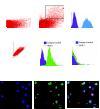
Labeling of human lung-resident mesenchymal stem (LR-MSCs) for in vivo tracking. (A) PKH-26 labeling of human lung–derived mesenchymal stem cells. Fluorescence microscopy (100× oil immersion) of cytospin of PKH-26–labeled LR-MSCs demonstrates homogenous red fluorescent membrane staining at Day 3 after labeling. A heterogeneous cytoplasmic distribution of PKH-26 was present by Day 7, consistent with internalization of labeled membrane. (B) DsRed labeling of human lung–derived mesenchymal stem cells. Histogram of human LR-MSCs transduced with control lentivirus, the pLentilox 3.7 vector containing DsRed sequences in its backbone, is shown. The transduction efficiency was 96.3 ± 2.9%, and cells demonstrated stable immunofluoresence with passaging.
Figure 2.
Figure 2.
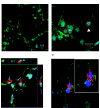
Retention of exogenously administered LR-MSCs in murine lungs. (A) Live PKH-26–positive cells can be identified in the murine lungs after exogenous administration of PKH-26–labeled human LR-MSCs. Flow cytometric analysis of single-cell suspension of lungs of SCID immunodeficient mice 3 weeks after intratracheal injection of saline or PKH-26–labeled human LR-MSCs is shown. Dot plot of propidium iodide–negative cells demonstrates a distinct live PKH26-positive population in lungs injected with labeled LR-MSCs (center panel). This is in contrast to cells from lungs of control saline–injected animals (left panel). A histogram of PKH-26–positive cells sorted from murine lungs is shown on the right. Control unlabeled LR-MSCs are shown in purple. (B and C) PKH-26–positive cells sorted from murine lungs are human in origin. (B) Dot plot (left panel) and histogram (center panel) demonstrating MHC class I expression on PKH-26–positive cells sorted by flow cytometry from murine lungs. The right panel demonstrates the absence of human MHC I expression on murine cells (mouse lung fibroblasts). Purple peaks represent isotype control. (C) Cytospin preparation of PKH-26–positive cells sorted from murine lungs demonstrates the presence of human nuclei on immunofluorescence staining using mouse antihuman nuclei monoclonal antibody.
Figure 3.
Figure 3.
LR-MSCs demonstrate superior retention in a murine lung. Dot plot of single-cell suspensions of murine lungs harvested 3 weeks after intratracheal administration of saline, DsRed-labeled LR-MSCs, and control cells (DsRed-labeled human foreskin and lung fibroblasts) are shown. A distinct population of DsRed-positive cells was noted in lungs injected with labeled human LR-MSCs as compared with the saline group. This population was significantly lower in lungs injected with labeled human foreskin or lung fibroblasts. Three weeks after intratracheal administration, 19.3 ± 3.21% of injected LR-MSCs, compared with 3.47 ± 0.51% of injected control foreskin fibroblasts and 4.82 ± 0.80% of injected control lung fibroblasts, were recovered at (n = 3 separate experiments of three animals each).
Figure 4.
Figure 4.
(A and B) Presence of exogenously administered human LR-MSCs in murine lungs at 6 months after intratracheal administration. At 6 months after intratracheal administration, LR-MSCs could still be distinctly identified in the alveolar spaces. Representative confocal microscopic images demonstrate LR-MSCs, identified by their red fluorescent PKH-26 staining. Mouse lung alveolar epithelial cells (AECs) are stained with cytokeratin (FITC, green) to better visualize the anatomy. LR-MSCs in the murine alveolar spaces demonstrated a round morphology with a prominent nucleus. LR-MSCs were seen predominantly near the corners of the alveoli embedded in the interstitium (arrow) or attached to the alveolar septa (solid arrow). Blue = DAPI (A: 20× magnification; B: 40× magnification). Bar = 10 μm. (C). Z stack imaging of confocal microscopy demonstrating PKH-26–labeled LR-MSCs embedded in the interstitium surrounded by AECs. Another LR-MSC (arrow) is seen attached to the alveolar septa near the corner lying adjacent to a type II AEC. (D) ZO-1 staining demonstrates the close proximity of an engrafted LR-MSC to type II AECs, with expression of ZO-1 (green) at the margin of these adjoining cells.
Figure 5.
Figure 5.
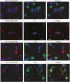
Exogenously administered human LR-MSCs maintain their mesenchymal phenotype but localize in close proximity to type II AECs. Immunofluorescent staining of murine lungs 6 months after intratracheal injection of PKH-26–labeled human LR-MSCs demonstrating their in situ phenotype in an alveolar microenvironment. PKH-26–labeled LR-MSCs (red) did not express surfactant protein (SP)C or thyroid transcription factor (TTF)-1 but were seen lying in close contact with SPC- or TTF-1–positive type II AECs. LR-MSCs resident in murine tissue continued to be negative for pan leukocyte marker CD45 and positive for cell surface glycoprotein receptor CD44. Bar = 10 μm.
Figure 6.
Figure 6.
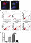
Demonstration of in vitro gap junction communication between human lung–resident MSCs and AECs. (A) Calcein AM–labeled LR-MSCs were cocultured with DiI-labeled human alveolar epithelial A549 cells for various time intervals in the absence or presence of the gap junction communication inhibitor carbenoxolone (CBX). The emergence of an increasing number of double-positive populations over time (circle) demonstrates transfer of Calcein from LR-MSCs to AECs. This transfer was inhibited in the presence of CBX (100 μM). (B) Quantitative analysis of dye transfer between LR-MSCs and AECs. Percentages of Calcein AM acceptor cells are shown over time. Each point represents the mean ± SD of three separate LR-MSC lines. *P < 0.0001. (C) Immunofluorescence staining of a coculture of LR-MSCs and AECs demonstrating expression of connexin 43. AECs can be identified by their cuboidal shape and cytokeratin staining (red). Connexin 43 staining is shown as green dots lining the spindle-shaped LR-MSCs. Bar = 10 μm.
Figure 7.
Figure 7.
Demonstration of in vivo gap junction communication between exogenously administered LR-MSCs and resident lung cells. (A) Connexin 43 expression in human LR-MSCs engrafted in murine lungs. Sections from murine lungs injected with human LR-MSCs were examined for expression of connexin 43. Connexin 43 (green) is seen as punctuate dots at the periphery of the engrafted PKH-26–stained LR-MSCs (red). Bar = 10 μm. (B) Demonstration of in vivo gap junction communications between LR-MSCs and tissue-resident cells. PKH-26–labeled and Calcein-labeled human LR-MSCs (1 × 10⁶) were injected intratracheally into murine (SCID) lungs. Flow cytometric analysis of single-cell homogenates of murine lungs at various time points after intratracheal injection demonstrates the emergence of a Calcein-AM–positive but PKH-26–negative population of acceptor cells (circle). This population was significantly decreased in the presence of CBX. (C) Quantitative analysis of dye transfer between LR-MSCs and murine lung resident cells. Percentage of Calcein AM–positive, PKH26–negative acceptor cells are shown over time. *P < 0.0001 (n = 4 separate experiments).

References

    1. Friedenstein AJ, Chailakhjan RK, Lalykina KS. The development of fibroblast colonies in monolayer cultures of guinea-pig bone marrow and spleen cells. Cell Tissue Kinet 1970;3:393–403 - PubMed
    1. Pittenger MF, Mackay AM, Beck SC, Jaiswal RK, Douglas R, Mosca JD, Moorman MA, Simonetti DW, Craig S, Marshak DR. Multilineage potential of adult human mesenchymal stem cells. Science 1999;284:143–147 - PubMed
    1. Dazzi F, Ramasamy R, Glennie S, Jones SP, Roberts I. The role of mesenchymal stem cells in haemopoiesis. Blood Rev 2006;20:161–171 - PubMed
    1. Brody AR, Salazar KD, Lankford SM. Mesenchymal stem cells modulate lung injury. Proc Am Thorac Soc 2010;7:130–133 - PMC - PubMed
    1. Tolar J, Le Blanc K, Keating A, Blazar BR. Concise review: hitting the right spot with mesenchymal stromal cells. Stem Cells 2010;28:1446–1455 - PMC - PubMed

Publication types

MeSH terms