. 2011 Mar 15;124(Pt 6):873-8.
doi: 10.1242/jcs.077396.

Phospho-regulation of kinesin-5 during anaphase spindle elongation

Affiliations

Phospho-regulation of kinesin-5 during anaphase spindle elongation

Rachel Avunie-Masala et al. J Cell Sci. .

Abstract

The kinesin-5 Saccharomyces cerevisiae homologue Cin8 is shown here to be differentially phosphorylated during late anaphase at Cdk1-specific sites located in its motor domain. Wild-type Cin8 binds to the early-anaphase spindles and detaches from the spindles at late anaphase, whereas the phosphorylation-deficient Cin8-3A mutant protein remains attached to a larger region of the spindle and spindle poles for prolonged periods. This localization of Cin8-3A causes faster spindle elongation and longer anaphase spindles, which have aberrant morphology. By contrast, the phospho-mimic Cin8-3D mutant exhibits reduced binding to the spindles. In the absence of the kinesin-5 homologue Kip1, cells expressing Cin8-3D exhibit spindle assembly defects and are not viable at 37°C as a result of spindle collapse. We propose that dephosphorylation of Cin8 promotes its binding to the spindle microtubules before the onset of anaphase. In mid to late anaphase, phosphorylation of Cin8 causes its detachment from the spindles, which reduces the spindle elongation rate and aids in maintaining spindle morphology.


Figures
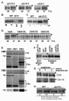
Fig. 1.
Fig. 1.
Cin8 is differentially phosphorylated during anaphase. Band-retardation assay in whole extracts of cells producing 6Myc–Cin8. Extracts were fractionated on SDS-PAGE and examined by Western blot. Arrowheads indicate the slow-migrating band of Cin8. (A) Cells expressing cdc15-2, cdc14-1 or cdc5-7 and containing 6Myc–Cin8 were grown at 26°C (permissive) and 37°C (restrictive). (B) Wild-type cells were either cycling or arrested at metaphase (by nocodazole), S-phase (by hydroxyurea) and G1 (by α-factor). (C) WT cells or cdc15-2 cells were grown at 37°C for 4 hours. The extracts were either treated with CIP phosphatase (pp) (+) or not (−). (D) cdc15-2 cells expressing either WT Cin8, Cin8-5A (S277, T285, S493, S736, S1010), Cin8-3A (S277, T285, S493) or Cin8-2A (S736, S1010) (indicated on top) were grown at 26°C or 37°C (indicated at the bottom). (E) In vitro phosphorylation of bacterially expressed Cin8-590 by Cdk1 using equal concentrations of the Cdk1 complexes with cyclin Clb2, Clb3 or Clb5 (indicated on top). Coomassie Brilliant Blue (CBB) staining and 32P autoradiograms of SDS-PAGE fractionation of phosphorylation reaction mixtures are shown (indicated on the left). Proteins corresponding to the various bands are indicated on the right, based on their predicted size. The relative specificity pattern obtained using equal concentrations of the kinase complexes with a reference substrate Histone H1 is shown in the bottom panel (H1). (F) Effect of CDC55 deletion on band-retardation assay of Cin8. Genotypes of cells and variants of Cin8–13Myc are indicated at the top; the various modes of arrest are indicated at the bottom. (G) Effect of Cdc14 overexpression on the slow-migrating band of Cin8–13Myc. 3HA–Cdc14 was produced from a galactose-inducible promoter. Cells were arrested either in S-phase or at late anaphase (indicated on top). In anaphase-arrested cells, Cdc14 was induced by addition of galactose for the times indicated above. The western blots of Cin8–13Myc (top) and 3HA–Cdc14 (bottom) are shown. (H) 6Myc–Cin8 was expressed in WT or spo12Δ cells (indicated on top). Cells were arrested in S-phase and released to fresh medium for 70 minutes (bottom).
Fig. 2.
Fig. 2.
Anaphase spindle localization of phosphorylation mutants of Cin8–3GFP. (A) a, Cin8; b, Cin8-2A, c, Cin8-3A, d, Cin8-3D. Time interval, 1 minute. (B) a, Cin8; b, Cin8-3A; time interval, 20 seconds. Scale bar: 1 μm. Arrows indicate spindle poles; arrowheads indicate the midzone.
Fig. 3.
Fig. 3.
Anaphase spindle elongation and morphology in cells that express phosphorylation mutants of Cin8. (A) Representative spindle-elongation kinetics in cells producing WT Cin8 (left), Cin8-3A (middle) and Cin8-3D (right). V, rate of spindle elongation (μm/minute). The phases of spindle elongation are indicated as follows: diamonds, fast phase; squares, slow phase; circles, post-anaphase. (B) Long anaphase spindle morphologies in cells expressing tubulin–GFP and Cin8 (top), Cin8-3A (middle) or Cin8-3D (bottom). Scale bar: 1 μm. Arrowheads indicate small overlapping region of the iMTs.

References

    1. Blangy A., Lane H. A., d'Herin P., Harper M., Kress M., Nigg E. A. (1995). Phosphorylation by p34cdc2 regulates spindle association of human Eg5, a kinesin-related motor essential for bipolar spindle formation in vivo. Cell 83, 1159-1169 - PubMed
    1. Buttery S. M., Yoshida S., Pellman D. (2007). Yeast formins Bni1 and Bnr1 utilize different modes of cortical interaction during the assembly of actin cables. Mol. Biol. Cell 18, 1826-1838 - PMC - PubMed
    1. Buvelot S., Tatsutani S. Y., Vermaak D., Biggins S. (2003). The budding yeast Ipl1/Aurora protein kinase regulates mitotic spindle disassembly. J. Cell Biol. 160, 329-339 - PMC - PubMed
    1. Cahu J., Olichon A., Hentrich C., Schek H., Drinjakovic J., Zhang C., Doherty-Kirby A., Lajoie G., Surrey T. (2008). Phosphorylation by Cdk1 increases the binding of Eg5 to microtubules in vitro and in Xenopus egg extract spindles. PLoS One 3, e3936 - PMC - PubMed
    1. Chee M. K., Haase S. B. (2010). B-cyclin/CDKs regulate mitotic spindle assembly by phosphorylating kinesins-5 in budding yeast. PLoS Genet. 6, e1000935 - PMC - PubMed

Publication types

MeSH terms