. 2011 Apr 20;30(8):1659-70.
doi: 10.1038/emboj.2011.60. Epub 2011 Mar 4.

A structural basis for Lowe syndrome caused by mutations in the Rab-binding domain of OCRL1

Affiliations

A structural basis for Lowe syndrome caused by mutations in the Rab-binding domain of OCRL1

Xiaomin Hou et al. EMBO J. .

Abstract

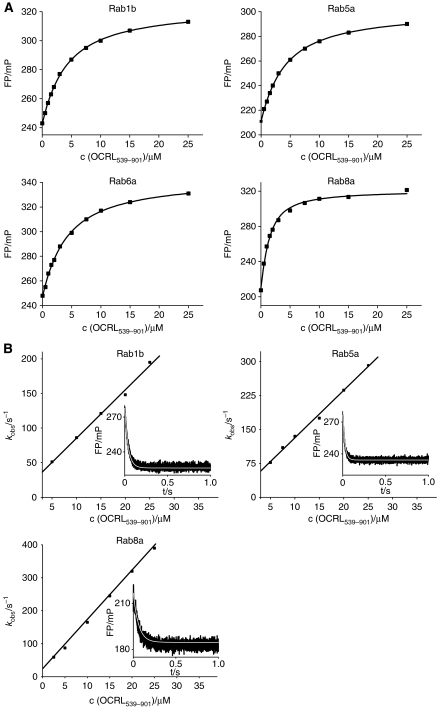
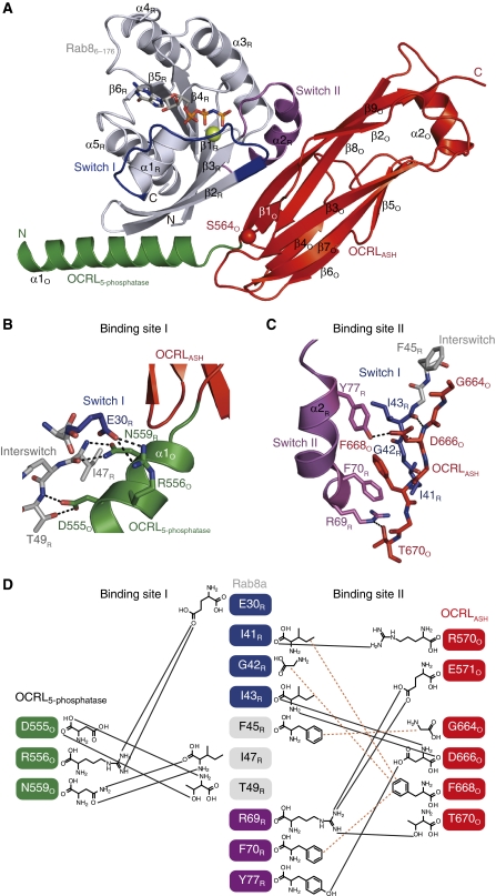
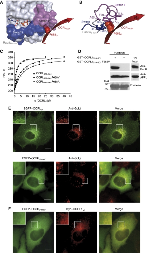
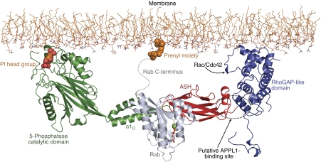
The oculocerebrorenal syndrome of Lowe (OCRL), also called Lowe syndrome, is characterized by defects of the nervous system, the eye and the kidney. Lowe syndrome is a monogenetic X-linked disease caused by mutations of the inositol-5-phosphatase OCRL1. OCRL1 is a membrane-bound protein recruited to membranes via interaction with a variety of Rab proteins. The structural and kinetic basis of OCRL1 for the recognition of several Rab proteins is unknown. In this study, we report the crystal structure of the Rab-binding domain (RBD) of OCRL1 in complex with Rab8a and the kinetic binding analysis of OCRL1 with several Rab GTPases (Rab1b, Rab5a, Rab6a and Rab8a). In contrast to other effectors that bind their respective Rab predominantly via α-helical structure elements, the Rab-binding interface of OCRL1 consists mainly of the IgG-like β-strand structure of the ASPM-SPD-2-Hydin domain as well as one α-helix. Our results give a deeper structural understanding of disease-causing mutations of OCRL1 affecting Rab binding.


Conflict of interest statement

The authors declare that they have no conflict of interest.

Figures

Figure 1
Figure 1
Schematic representation of the domain architecture of OCRL1.
Figure 2
Figure 2
OCRL1 binds different exocytic and endocytic Rab proteins with moderate affinities. (A) Fluorescence polarization based equilibrium titration of 1 μM Rab1b:mantGppNHp, Rab5a:mantGppNHp, Rab6a:mantGppNHp or Rab8a:mantGppNHp with OCRL1539–901, respectively. (B) Association and dissociation kinetics of Rab1b:mantGppNHP, Rab5a:mantGppNHp and Rab8a:mantGppNHp with OCRL1539–901, respectively, monitored by the change in fluorescence polarization using stopped-flow apparatus (c(RabX:mantGppNHp)=1 μM). The individual observed association rate constants are linearly dependent on the OCRL1539–901 concentration and were fitted to a straight line, yielding the association rate constant. (Insets) The dissociation of fluorescent RabX:mantGppNHp (2 μM) from its complex with OCRL1539–901 was monitored by displacement with a 10-fold molar excess of non-fluorescent Rab using the stopped-flow apparatus. The time-dependent decrease in fluorescence polarization, indicative of complex dissociation, was fitted to a single exponential function (white line) to determine the dissociation rate constants. FP, fluorescence polarization; mP, milli-polarization units.
Figure 3
Figure 3
Crystal structure of the Rab8a6–176:OCRL1540–678 complex (A) Cartoon representation of the Rab8a6–176:OCRL1540–678 complex. Rab8a6–176 is shown in grey, with Switch I in blue and Switch II in magenta. OCRL1540–670 is in green (helix α1O of the 5-phosphatase domain) and red (ASH domain). The position of S564O (S564PO causes Lowe syndrome) is indicated with a red sphere. (N, N-termini; C, C-termini; sticks, non-hydrolyzable GTP-analogue GppNHp; green sphere, Mg2+, colouring is kept consistent throughout all panels). (B, C) Detailed view of interactions between Rab8a6–176 with α1O-helix (binding site I, (B)) and the ASH domain (binding site II, (C)) of OCRL1540–678. The Switch I region of Rab8a and the β9O-strand of OCRL1 are in stick representation to show the main-chain interactions (black dashes: polar interactions). (D) Schematic view of Rab8a–OCRL1 interactions (black lines: hydrogen bonds, brown dashes: hydrophobic contacts).
Figure 4
Figure 4
The binding of APPL1 and Ses1 peptides does not alter the dissociation equilibrium constant of the Rab8:mantGppNHp–OCRL1539–901 interaction. The experimental setup described in Figure 2A was repeated in the presence of 200 μM APPL1 peptide or 100 μM Ses1 peptide with 1 μM Rab8a:mantGppNHp and varying concentrations of OCRL1539–901 (FP, fluorescence polarization; mP, milli-polarization units).
Figure 5
Figure 5
Structural and thermodynamic basis for the Lowe syndrome causing mutation F668V. (A) The residue F668O (red spheres) of the β9O-strand of OCRL1 (red cartoon) is binding within a hydrophobic pocket of Rab8a (surface representation) located between Switch I and Switch II (the nucleotide is shown as ball and stick representation). (B) F668O is penetrating into a hydrophobic pocket of seven hydrophobic residues (I41R, G42R, I43R, A65R, F70R, I73R and Y77R). (C) Fluorescence polarization based equilibrium titrations of Rab8a:mantGppNHp with different OCRL1539–901 variants (wild type, F668VO and F668AO), demonstrating the significance of the Lowe disease related mutant F668O for Rab binding. The mutations F668VO and F668AO of OCRL1 decrease the affinity to Rab8a (KD,Rab8:OCRL1 wt=0.9 μM, KD,Rab8:OCRL1 F668V=5.2 μM, KD,Rab8:OCRL1 F668A=6.0 μM). (D) Pull-down experiment of Rab8a with GST–OCRL1 fusion proteins. Indicated GST-fusion proteins were incubated with Hela cell lysate, bound proteins were separated by SDS–polyacrylamide gel electrophoresis transferred to nitrocellulose and detected using anti-Rab8a or anti-APPL1 antibody, equal loading of GST-fusion proteins is indicated by ponceau staining. (E) Subcellular localization of OCRL1 F668V. Hela cells expressing GFP-tagged full-length OCRL1 or OCRL1 point mutant F668V were analysed by immunofluorescence microscopy with antibodies to the Golgi 58K protein. Insets show an enlargement of the Golgi area. (F) Comparison of subcellular localizations of OCRL1 wt with OCRL1 F668V. Hela cells were co-transfected with constructs for EGFP–OCRL1 F668V and Myc-tagged wild-type OCRL1 and analysed using immunofluorescence microscopy with antibodies against the Myc epitope (Bar, 10 μm).
Figure 6
Figure 6
Structural model of the interaction of OCRL1 and Rab8a at the membrane interface. The inositol-5-phosphatase domain (Tsujishita et al, 2001) together with the ASH and RhoGAP-like domains (Erdmann et al, 2007) are docked at the membrane interface as reported previously. The N-terminal region of OCRL1 is not included. The Rab protein binds centrally between the 5-phosphatase and RhoGAP domains, with the nucleotide-binding pocket (sticks, GppNHp; green sphere, Mg2+) facing towards the cytosol. In this model, binding of Rac/Cdc42 to the RhoGAP domain and APPL1 to OCRL1 (dashed circle) simultaneously with Rab8a is conceivable.

References

    1. Attree O, Olivos IM, Okabe I, Bailey LC, Nelson DL, Lewis RA, McInnes RR, Nussbaum RL (1992) The Lowe Oculocerebrorenal Syndrome Gene Encodes a Protein Highly Homologous to Inositol Polyphosphate-5-Phosphatase. Nature 358: 239–242 - PubMed
    1. Berrow NS, Alderton D, Sainsbury S, Nettleship J, Assenberg R, Rahman N, Stuart DI, Owens RJ (2007) A versatile ligation-independent cloning method suitable for high-throughput expression screening applications. Nucleic Acids Res 35: e45. - PMC - PubMed
    1. Bleimling N, Alexandrov K, Goody R, Itzen A (2009) Chaperone-assisted production of active human Rab8A GTPase in Escherichia coli. Protein Expr Purif 65: 190–195 - PubMed
    1. Burguete AS, Fenn TD, Brunger AT, Pfeffer SR (2008) Rab and Arl GTPase family members cooperate in the localization of the golgin GCC185. Cell 132: 286–298 - PMC - PubMed
    1. Chavas LM, Ihara K, Kawasaki M, Torii S, Uejima T, Kato R, Izumi T, Wakatsuki S (2008) Elucidation of Rab27 recruitment by its effectors: structure of Rab27a bound to Exophilin4/Slp2-a. Structure 16: 1468–1477 - PubMed

Publication types

MeSH terms