. 2011 Feb 17;6(2):e16539.
doi: 10.1371/journal.pone.0016539.

Characterisation of the putative effector interaction site of the regulatory HbpR protein from Pseudomonas azelaica by site-directed mutagenesis

Affiliations

Characterisation of the putative effector interaction site of the regulatory HbpR protein from Pseudomonas azelaica by site-directed mutagenesis

Christelle Vogne et al. PLoS One. .

Abstract

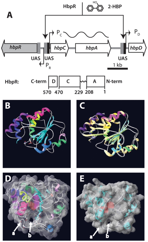
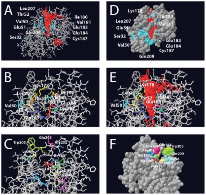
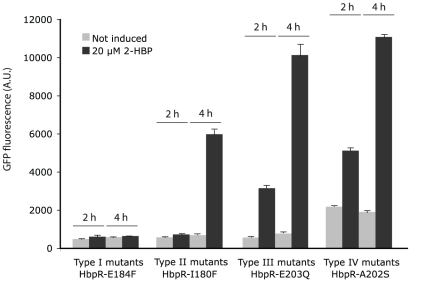
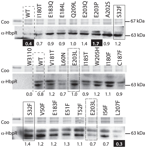
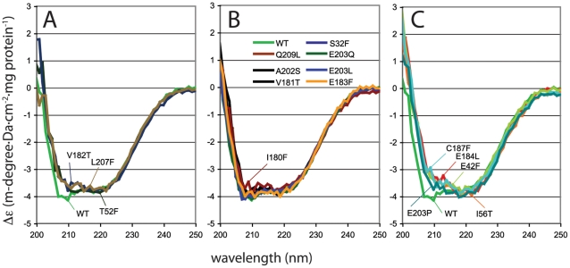
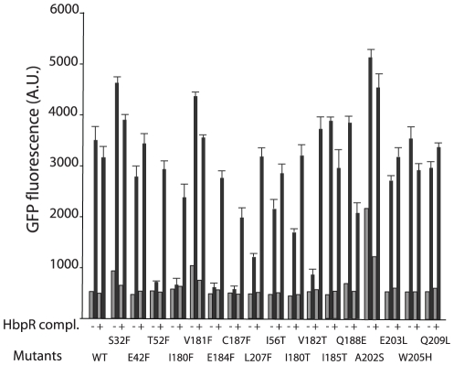
Bacterial transcription activators of the XylR/DmpR subfamily exert their expression control via σ(54)-dependent RNA polymerase upon stimulation by a chemical effector, typically an aromatic compound. Where the chemical effector interacts with the transcription regulator protein to achieve activation is still largely unknown. Here we focus on the HbpR protein from Pseudomonas azelaica, which is a member of the XylR/DmpR subfamily and responds to biaromatic effectors such as 2-hydroxybiphenyl. We use protein structure modeling to predict folding of the effector recognition domain of HbpR and molecular docking to identify the region where 2-hydroxybiphenyl may interact with HbpR. A large number of site-directed HbpR mutants of residues in- and outside the predicted interaction area was created and their potential to induce reporter gene expression in Escherichia coli from the cognate P(C) promoter upon activation with 2-hydroxybiphenyl was studied. Mutant proteins were purified to study their conformation. Critical residues for effector stimulation indeed grouped near the predicted area, some of which are conserved among XylR/DmpR subfamily members in spite of displaying different effector specificities. This suggests that they are important for the process of effector activation, but not necessarily for effector specificity recognition.


Conflict of interest statement

Competing Interests: The authors have declared that no competing interests exist.

Figures

Figure 1
Figure 1. Overview of the hbp regulatory system and tertiary structure modeling of the HbpR A-domain using a previously established XylR model as template.
(A) Organization of the hbp genes and the location of the HbpR binding sites (UAS, upstream activating sequences) in front of the PC and PD promoters. HbpR domains are depicted to scale according to the predictions by Jaspers et al . (B) to (E) Fitting used swiss-model and was performed on XylR A-domain PDB coordinates as calculated by Devos et al . (B) Ribbon model of HbpR A-domain residues 11–209, with predicted coils, alpha-helices and beta-sheets indicated. (C) Superposition of the predicted HbpR and XylR A-domains in the same configuration as A. (D) Tertiary structure model of HbpR A-domain with calculated molecular surface at 1.4Å and 40% transparency, in order to see the helical, coil and sheets. Model turned into a position which enables visualization of the proposed tunnel entry (b). C-terminal end of coil ending the A-domain indicated with an arrow at (a). Pinkish region in the centre of the A-domain illustrates a predicted cavity within the A-domain. (E), as C but now for the XylR A-domain, with exception of the ten most C-terminal residues, which otherwise are predicted to occlude the tunnel.
Figure 2
Figure 2. Details of the modeled tertiary structure of the HbpR A-domain, showing amino acid residues that were mutated in this study and the region onto which 2-HBP is predicted to be bound.
(A) Results of 1000 iterations of 2-HBP (in red) docking calculations using gramm onto the predicted HbpR A-domain protein surface, whilst indicating the position of residues altered to Phe. (B) Close-up of the same, but without the docked 2-HBP positions. (C) as for B, now highlighting the other changed residues. (D) Van der Waals-filled model slightly turned compared to A, in order to indicate the region of 2-HBP docked molecules. (E), as B, but with 2-HBP docked positions. (F) Turned van der Waals-filled model showing the tunnel from the other side of the entry.
Figure 3
Figure 3. Exemplary effects of HbpR A-domain mutations on inducible expression.
Measured fluorescence intensities of Escherichia coli cells carrying a plasmid with a promoterless egfp under control of the HbpR-dependent PC-promoter in the presence or absence of 20 µM 2-HBP as inducer. EGFP expression was measured on whole cells at two time points and corrected for culture turbidity. Type I to IV correspond to differently shaded entries in Table 1. Note the delayed response in Type II mutants and the higher background in the absence of inducer in type IV mutants.
Figure 4
Figure 4. HbpR (mutant) expression in E. coli from pHBP269A0-plasmids, i.e., those which were used for 2-HBP induced EGFP expression from PC. Coo, Coomassie-Blue-stained SDS-PAGE gel fragment around 67 kDa. α-HbpR, bands on gel detected in Western blotting with anti-HbpR antibodies.
Relevant mutations are indicated; note that wild-type and several mutants were analyzed twice. Numbers below the gel fragments indicate the average normalized intensities of both HbpR bands for each mutant or wild-type. Those numbers highlighted in white on black background point to values below or above the 25 and 75% quantiles of all measured intensities.
Figure 5
Figure 5. Circular dichroism spectra of purified His6-tagged HbpR wild-type protein or of sixteen purified HbpR A-domain mutants, between 200 and 250 nm, at a protein concentration of ≈0.3 mg/ml.
Spectra were normalized to Δε, as indicated in the Experimental Procedures section, and grouped to reveal similar dichroism trends. (A) HbpR wild-type and mutants V182T, L207F and T52F (type II effects with delayed and lower induction by 2-HBP). (B) Mutants with similar dichroisms as HbpR wild-type. (C) Mutants with the most strong aberration of the wild-type circular dichroism trace, of which C187F and E184L completely abolished activation by 2-HBP, but E42F and E203P having no major effect on 2-HBP dependent induction in E. coli.
Figure 6
Figure 6. EGFP expression from the HbpR-dependent PC-promoter in E. coli in the presence (black bars) or absence (grey bars) of 2-HBP as inducer measured for the different HbpR A-domain mutants after 2 h induction time, and either complemented with a second plasmid carrying wild-type HbpR (pHB240) or not.
Note the partial ‘rescue’ of the abolished phenotype in T52F, I180F, E184F, C187F and V182T by wild-type HbpR, suggesting that the mutant proteins are not dominantly negative over the wild-type. Results indicate the mean of triplicate incubations, plus the calculated standard deviation.

References

    1. Shingler V. Signal sensing by σ54-dependent regulators: derepression as a control mechanism. Mol Microbiol. 1996;19:409–416. - PubMed
    1. Tropel D, van der Meer JR. Bacterial transcriptional regulators for degradation pathways of aromatic compounds. Microbiol Mol Biol Rev. 2004;68:474–500. - PMC - PubMed
    1. Ramos JL, Marques S, Timmis KN. Transcriptional control of the Pseudomonas TOL plasmid catabolic operons is achieved through an interplay of host factors and plasmid- encoded regulators. Annu Rev Microbiol. 1997;51:341–373. - PubMed
    1. Shingler V, Moore T. Sensing of aromatic compounds by the DmpR transcriptional activator of phenol-catabolizing Pseudomonas sp. strain CF600. J Bacteriol. 1994;176:1555–1560. - PMC - PubMed
    1. Arenghi FL, Barbieri P, Bertoni G, de Lorenzo V. New insights into the activation of o-xylene biodegradation in Pseudomonas stutzeri OX1 by pathway substrates. EMBO Rep. 2001;2:409–414. - PMC - PubMed

Publication types

MeSH terms

Substances