. 2011 Aug;66(2):314-23.
doi: 10.1002/mrm.22805. Epub 2011 Mar 4.

Real-time motion and B0 corrected single voxel spectroscopy using volumetric navigators

Affiliations

Real-time motion and B0 corrected single voxel spectroscopy using volumetric navigators

Aaron T Hess et al. Magn Reson Med. 2011 Aug.

Abstract

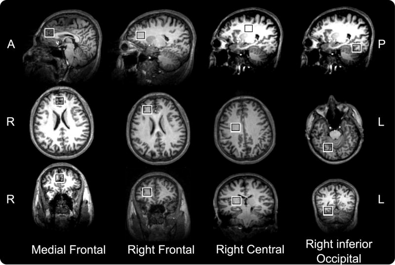
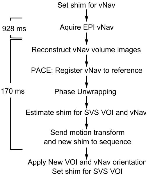
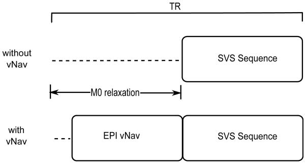
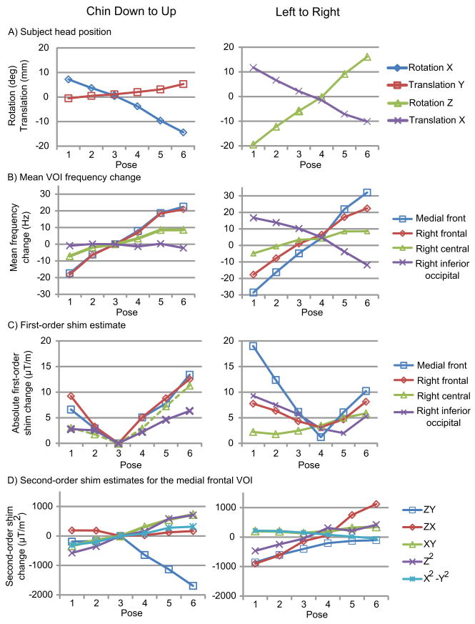
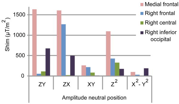
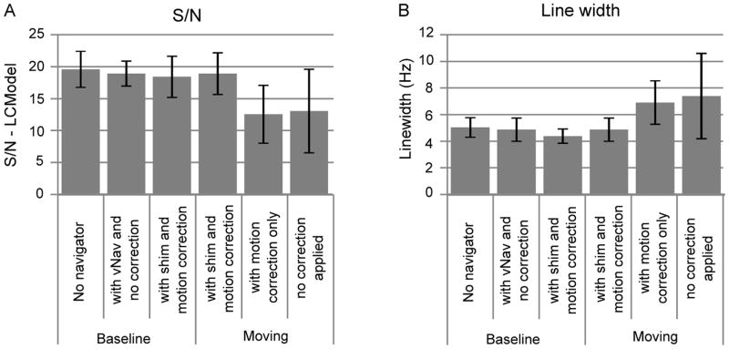
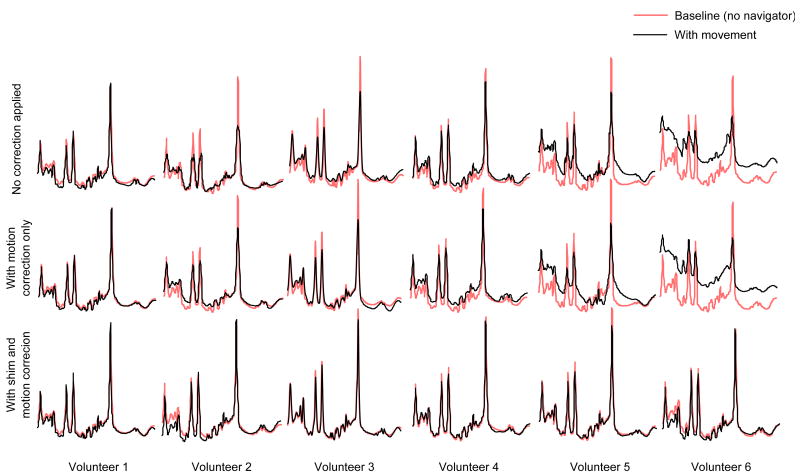
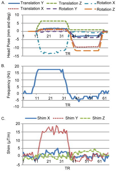
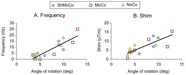
In population groups where head pose cannot be assumed to be constant during a magnetic resonance spectroscopy examination or in difficult-to-shim regions of the brain, real-time volume of interest, frequency, and shim optimization may be necessary. We investigate the effect of pose change on the B0 homogeneity of a (2 cm)3 volume and observe typical first-order shim changes of 1 μT/m per 1° rotation (chin down to up) in four different volumes of interest in a single volunteer. An echo planar imaging volume navigator was constructed to measure and apply in real-time within each pulse repetition time: volume of interest positioning, frequency adjustment, and first-order shim adjustment. This volume navigator is demonstrated in six healthy volunteers and achieved a mean linewidth of 4.4 Hz, similar to that obtained by manual shim adjustment of 4.9 Hz. Furthermore, this linewidth is maintained by the volume navigator at 4.9 Hz in the presence of pose change. By comparison, a mean linewidth of 7.5 Hz was observed, when no correction was applied.


Figures

Figure 1
Figure 1
A. Change in linewidth as a function of first-order B0 inhomogeneity for 40 ms, 80 ms and 160 ms inherent linewidths in a (2cm)3 voxel. B. Change in linewidth as a function of first-order B0 inhomogeneity for varying voxel sizes, (1 cm)3, (2 cm)3, and (3 cm)3. C. Change in linewidth against second-order B0 gradient for ZX, ZY, XY, Z2 and (X2 – Y2) at an inherent linewidth of 80 ms. D. Spectral amplitude as a function of second-order B0 terms ZX, ZY, XY, Z2 and (X2 – Y2), relative to the amplitude in a homogeneous VOI.
Figure 2
Figure 2
VOI's Medial Frontal, Right Frontal, Right Central, and Right inferior Occipital, for which change in B0 shim gradients with respect to movement are demonstrated.
Figure 3
Figure 3
Work flow of vNav block, sequence and online processing
Figure 4
Figure 4
A typical SVS PRESS sequence and our navigated SVS PRESS with vNav inserted into the M0 relaxation period.
Figure 5
Figure 5
B0 changes as a result of chin down - up and left - right motion. A. Motion trajectory, B. Mean VOI frequency change for each VOI, C. Absolute magnitude of first-order B0 shim vector independent of second-order for each VOI, and D. Second-Order B0 shim estimates for the Medial Frontal VOI, offset by the value at rest. X, Y, and Z refer to the scanner axes perpendicular to the sagittal, coronal, and transverse planes and the X axis labels 1 to 6 refer to each of six respective head positions.
Figure 6
Figure 6
Magnitude of the second-order B0 terms in the neutral pose compared for each of the four VOI's demonstrate that the frontal lobe has the highest second-order shim requirements.
Figure 7
Figure 7
Example navigator volumes. A. Magnitude images for first echo, and B. Unwrapped and masked field map with the contrast range doubled (-2π to 2π) due to the phase unwrapping.
Figure 8
Figure 8
A. Bar graph of the mean signal to noise ratio (S/N as calculated by LCModel, +/- one standard deviation). B. Bar graph of the mean linewidth (+/- one standard deviation). Both calculated over the 6 volunteers for each of the three stationary baseline scans and each of the three acquisitions with motion.
Figure 9
Figure 9
Spectra obtained in the right central white matter for the three scans acquired with movement (no correction, with motion correction, and with full shim and motion correction) for all six volunteers superimposed on top of the respective baseline spectra with no navigator. The plots are the spectra as fitted by LCModel.
Figure 10
Figure 10
Navigator output and frequency variation from the motion corrected acquisition of volunteer 6. A. Absolute motion estimate as calculated by the navigator, B. mean VOI frequency as calculated from FID cross-correlation, and C. first-order B0 shim change as calculated by the navigator, all as a function of the TR over the duration of the acquisition. X, Y, and Z refer to the scanner axes perpendicular to the sagittal, coronal, and transverse planes, respectively.
Figure 11
Figure 11
A. Scatter plot of change in frequency and B. change in Y shim gradient as a function of the angle of chin-up rotation about X for all three scans with motion from the six volunteers as measured by the vNav. These values were calculated form the maximum chin-up rotation, averaged over the duration that the subject maintained that pose.

References

    1. Ernst T, J L. Phase navigators for localized MR spectroscopy using water suppression cycling. Proceedings of the 17th annual meeting of the ISMRM; Honolulu, HI. 2009. p. 239.
    1. Helms G, Piringer A. Restoration of motion-related signal loss and line-shape deterioration of proton MR spectra using the residual water as intrinsic reference. Magnetic Resonance in Medicine. 2001;46(2):395–400. - PubMed
    1. Posse S, Cuenod CA, Le Bihan D. Motion artifact compensation in 1H spectroscopic imaging by signal tracking. Journal of Magnetic Resonance Series B. 1993;102:222.
    1. Star-Lack JM, Adalsteinsson E, Gold GE, Ikeda DM, Spielman DM. Motion correction and lipid suppression for 1H magnetic resonance spectroscopy. Magnetic Resonance in Medicine. 2000;43(3):325–30. - PubMed
    1. Henry PG, van de Moortele PF, Giacomini E, Nauerth A, Bloch G. Field-frequency locked in vivo proton MRS on a whole-body spectrometer. Magnetic Resonance in Medicine. 1999;42(4):636–42. - PubMed

Publication types

MeSH terms